Other Endocrine Glands
Parathyroid Glands
The parathyroid glands are small aggregates of endocrine tissue within or near the thyroid
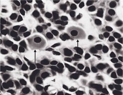
Figure 12-9.
An equine parathyroid gland with two large oxyphil cells (arrows) among numerous chief cells. (Reprinted with permission of Wiley-Blackwell from Dellmann H.D. Textbook of Veterinary Histology. 5th ed. Baltimore: Lippincott Williams & Wilkins, 1998.)gland. Most domestic animals have two pairs of parathyroid glands, but the exact number and location vary with the species. Commonly, one pair is visible outside the thyroid gland and is therefore designated external parathyroids. The second pair is often buried in the substance of the thyroid, and these are the internal parathyroids. The pig lacks grossly visible internal parathyroids.
The two types of parathyroid cells are chief cells and oxyphil cells (Fig. 12-9). Chief cells are small, usually dark cells that are associated with the production of parathyroid hormone (PTH, parathormone). The less numerous oxyphil cells are larger, with granular cytoplasm and a small, dark nucleus. These cells have been described in horses and cattle (and humans) but are not found in other domestic species. Their function is unknown, although the fact that they appear later in life has suggested that they may be senescent chief cells.
Parathyroid Hormone. Parathyroid hormone (PTH), a peptide hormone, is the major controller of the level of blood calcium and phosphate. it does this by stimulating release of calcium and phosphate from bone, decreasing excretion of calcium and increasing excretion of phosphate by the kidney, and promoting the formation of active vitamin D by the kidney (discussed later). In bone, PTH acts on both osteocytes and osteoclasts to promote the release of calcium phosphate.
The action of PTH on osteocytes releases calcium and phosphate from stores associated with the extracellular fluids in and around bone. PTH stimulates the degradation of bone by osteoclasts while it inhibits bone formation by osteoblasts. The overall effect of PTH is to increase blood calcium concentration and lower blood phosphate concentration by increasing its urinary excretion.The forms of vitamin D consumed in the diet or produced in the skin by the action of ultraviolet light on precursors is not the most active form of the vitamin. These forms are further metabolized in the liver to a second precursor form that is further metabolized in the kidneys to the most biologically active form. It is this final form (1,25-dihydroxycholcalcif- erol or calcitriol) that functions as the true vitamin D. PTH promotes the formation of this final metabolite within the kidney. Two major functions of vitamin D are to increase the rate of calcium absorption from the gastrointestinal tract and to reduce the loss of calcium in the urine. Thus, the overall effect of vitamin D is to retain calcium in the body.
The only significant regulator of PTH release is the concentration of ionized calcium in the blood plasma. Normally, about 50% of plasma calcium is bound to plasma proteins (primarily albumin), and this bound calcium is not biologically active. The other 50% is unbound and exists as calcium ions. chief cells detect decreases in the concentration of ionized calcium, and they respond by increasing their secretion of PTH. The resulting rise in plasma calcium has a negative feedback effect on further PTH secretion.
Diets insufficient in calcium are uncommon in domestic animals other than carnivores, but they can occur, especially with diets formulated primarily on grain products, which typically have high phosphorus and low calcium. In such cases, the chronically low intake of dietary calcium stimulates increased secretion of PTH to keep blood calcium levels adequate for nerve and muscle function.
Calcium is removed from the bone matrix, and this decalcification can lead to bone deformities and osteoporosis. This creates the medical condition nutritional secondary hyperparathyroidism. In young animals, growth may be disrupted and limb and spine abnormalities may develop, a manifestation of hyperparathyroidism sometimes called rickets. A particular form of nutritional hyperparathyroidism commonly called bran disease or bighead is seen in horses fed excessive amounts of cereal by-products (e.g., bran) in conjunction with calcium-poor hay. Bone tissue that has been decalcified by high levels of circulating PTH is frequently replaced with soft, bulky fibrous tissue; this is especially prominent in the flat bones of the face and mandible, giving rise to the appearance of an enlarged head. Prompt correction of the dietary imbalance may improve most symptoms (e.g., lameness) of bran disease, although the enlarged facial features are usually permanent.Pancreatic Islets
The pancreas of domestic animals is a bilobed gland adjacent to the proximal part of the duodenum (small intestine). The pancreas is an important exocrine gland whose enzymatic secretions are delivered to the lumen of the duodenum by one or two ducts. Scattered throughout the substance of the pancreas are small masses of endocrine tissue called pancreatic islets (formerly the islets of Langerhans).
The pancreatic islets are clumps of palestaining cells, arranged in irregular cords separated by capillaries (Fig. 12-10). Special stains are used to demonstrate the types of epithelial cells found in the pancreatic islets. The known number of distinct kinds of cells is still growing, but the two best characterized are the α-cells and β-cells. The β-cells are the more numerous (about 75% of all cells in the islet), and they produce the hormone insulin. α-Cells produce the hormone glucagon.
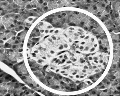
Figure 12-10.
Pancreatic islet (encircled) surrounded by cells of exocrine pancreas. (Courtesy of Sandra Pit- caithley, DVM.)β-Cells are sensitive to increases in blood glucose (sugar), such as occur after a meal containing digestible carbohydrates, and they release insulin in response to such increases in blood glucose. insulin lowers blood glucose by stimulating the uptake of glucose by many cells of the body, including skeletal muscle. insulin also stimulates skeletal muscle and liver cells to synthesize glycogen, the storage form of glucose. It affects the metabolism of amino acids and lipids, for it stimulates protein synthesis in skeletal muscle and liver and the deposition of lipids in adipose tissue. insulin is the major endocrine stimulus for the state of anabolism that exists after a meal is digested and nutrients are absorbed. When blood glucose decreases (such as during fasting), the stimulus for insulin secretion is lost and insulin levels are extremely low.
Glucagon causes liver cells to break down glycogen to release glucose, stimulates adipocytes to release fatty acids, and increases the synthesis of glucose in the liver from substrates other than carbohydrates, such as amino acids. The stimulus for glucagon release is a decrease in blood glucose to levels associated with fasting. α-Cells detect such decreases and respond by secreting glucagon in proportion to the reduction in blood glucose.
Insulin is necessary for the uptake of glucose by many cells, including skeletal muscle, which makes up most of the body mass. Without sufficient insulin, glucose accumulates in the blood after a meal, for it cannot be transported across cell membranes into cells where it can be used for fuel. The metabolic consequences of insufficient insulin (or of resistance to its effect) create the condition called diabetes mellitus.
Epiphysis (Pineal Gland)
The epiphysis cerebri (pineal gland or pineal body) is a midline structure on the dorsocaudal aspect of the diencephalon.
In fish, amphibians, and some reptiles, it possesses photoreceptors, and its proximity to the thin calvaria makes it literally a third eye, the function of which is thought to involve setting daily and yearly biologic cycles based on photoperiod. In mammals, including humans and domestic animals, the pineal has no photoreceptors, and its location deep inside the braincase renders it incapable of detecting photoperiods directly. The epiphysis nonetheless does receive information about light and dark cycles indirectly from a nucleus of the hypothalamus. The cells of the epiphysis, although neuronal by lineage, are secretory, and they are supported by neuroglia and receive axonal input. These specialized cells are called pinealocytes.The pinealocytes manufacture serotonin and an enzyme that converts this peptide to melatonin, a hormone. Manufacture of melatonin exhibits a profound diurnal rhythm, with releases into the blood peaking during darkness. It is likely, therefore, to be intimately involved in regulating sleep-wake cycles. It appears, too, to be linked to reproduction, inasmuch as the onset of puberty is associated with a profound fall in production of melatonin.
Early discoveries about the link between melatonin and sleep and reproduction have spawned a wave of enthusiastic speculation about its possible use as a cure for insomnia, an aphrodisiac, a treatment for jet lag, and an antiaging drug, among others. To date, however, no controlled studies have supported any of these claims.