Ovaries
The ovaries, like the testes in the male, are the primary organs of reproduction in the female. The ovaries are both endocrine and cytogenic (cell producing) as they produce hormones, which are released directly into the blood stream, and ova, which are released from the surface of the ovary during ovulation (see Chapter 27).
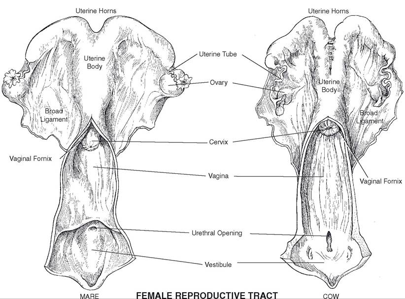
Figure 26-1.
Anatomy of the female reproductive tract.The ovaries are paired glands usually found in the lumbar region of the abdominal cavity, a short distance caudal to the kidneys. Like all abdominal organs, the ovaries are covered with peritoneum. They are suspended from the body wall by a reflection of this serous membrane, the mesovarium, the most cranial part of the peritoneal investments of the female genital tract.
in most species, the ovaries are somewhat ovoid (Fig. 26-2). In the mare, however, the ovaries have a bean shape because of a definite ovulation fossa, an indentation in the attached border of the ovary. ovaries of the sow usually appear lobulated because of the numerous developing ova; the sow is the only common farm animal that typically produces a litter, rather than one or two offspring per pregnancy.
When palpated through the wall of the rectum, an ovary feels solid because of the large amount of connective tissue that makes up the stroma of the gland. Normal size of the ovary varies considerably from species to species, and even within a species there is some variation. For example, the ovary of the mare may be less than 2.5 cm in diameter when no developing ova are present or as large as 10 cm with many developing ova.
The ovary is invested in a dense connective tissue capsule, the tunica albuginea. The medulla, or central portion, of the ovary is the most vascular part, while the cortex, or outer portion, consists largely of dense, irregular connective tissue interspersed with follicles (developing ova; see Fig.
27-1) and interstitial cells, which have an endocrine function.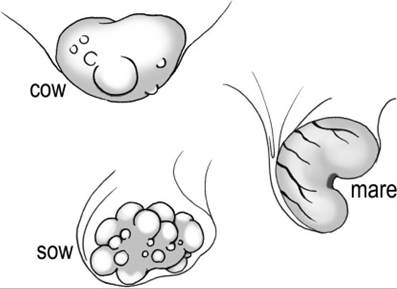
Figure 26-2. Ovaries of various species. Note the ovulation fossa on the equine ovary.
| Table 26-1. Comparative Anatomy of the Reproductive Tract in the Adult Nonpregnant Female | ||||
| Organ | Animal | |||
| Cow | Ewe | Sow | Mare | |
| Uterine tubea | 25 | 15-19 | 15-30 | 20-30 |
| Uterus | ||||
| Type | bi partiteb | bipartite | bicornuatec | bipartite |
| Horna | 35-40 | 10-12 | 40-65 | 15-25 |
| Bodya | 2-4 | 1-2 | 5 | 15-20 |
| Endometrium | 70-120 caruncles | 88-96 caruncles | Slight longitudinal folds | Conspicuous longitudinal folds |
| Cervix | ||||
| Lumen | 2-5 annular rings | annular rings | corkscrewlike | conspicuous folds |
| Opening to uterus | small and protruding | small and protruding | ill-defined | clearly defined |
| Vaginaa | 25-30 | 10-14 | 10-15 | 20-35 |
| Vestibulea | 10-12 | 2.5-3 | 6-8 | 10-12 |
aLength in centimeters. bBody divided into two parts. cUterus dominated by two horns.