Uterine Tubes
The uterine tubes (also called oviducts) are paired, convoluted tubes that conduct the ova from each ovary to the respective horn of the uterus (Fig. 26-1) and are the usual site of fertilization of ova by the spermatozoa.
The portion of the uterine tube adjacent to the ovary is expanded to form a funnel-shaped infundibulum. The infundibulum appears to take an active part in ovulation, at least to the extent of partially or completely enclosing the ovary and directing the ovum into the uterine tube.The lining of the uterine tube is a much- folded mucous membrane covered primarily with a simple columnar ciliated epithelium (Fig. 26-3). During estrus (period of sexual receptivity), the unciliated cells become actively secretory. The rest of the wall of the uterine tube includes a connective tissue submucosa and a muscular layer of smooth muscle. Both cilia and muscles function in the movement of ova and possibly in the movement of spermatozoa. The uterine tube, like the entire genital tract, is invested externally with peritoneum, which reflects off the organ as a suspending mesentery. The portion supporting the uterine tube is the mesosalpinx.
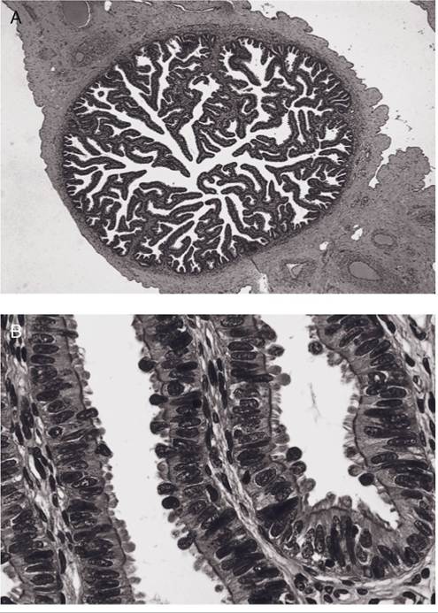
Figure 26-3. Histology of the uterine tube. A) Low-power micrograph in the region of the infundibulum. Note the elaborate folding of the epithelium. B) Higher-power micrograph showing the ciliated simple columnar epithelium of the uterine tube. The mucosal surface is characterized by abundant secretory product. (Reprinted with permission of Wiley-Blackwell from Bacha, Jr. W.J. and Bacha L.M. Color Atlas of Veterinary Histology. 2nd ed. Baltimore: Lippincott Williams & Wilkins, 2000.)