Overview of Function and Histology of the Kidneys
The urinary system is responsible for maintaining the relatively constant internal environment of the body fluids. This is accomplished by the formation and excretion of urine of an appropriate volume and composition.
Urine formation occurs in the kidneys, and by adjusting the volume and composition of urine in response to changes in dietary intake or metabolism, the kidneys regulate the body balance of water, various electrolytes, acids, and bases. The kidneys also excrete metabolic waste products in the urine, including the nitrogenous waste, urea, and a by-product of skeletal muscle metabolism, creatinine. Signs of kidney diseases include imbalances of water, electrolytes, acids, and bases and increases in blood levels of urea and creatinine.Kidneys are composite organs that consist of thousands to millions of similar microscopic functional units, the nephrons. Nephrons in all mammalian kidneys are similar in basic structure and function, but the number of nephrons differs among mammals. Large animals have more nephrons per kidney than small animals (e.g., 4 million for cattle and 500,000 for dogs). Nephrons consist of a spherical structure (Bowmans capsule) that contains a capillary tuft (glomerulus) and a single long tubule connected to Bowman’s capsule (Fig. 23-4). Bowman’s capsule consists of two layers of cells. The inner (visceral) layer closely surrounds the glomerular capillaries, and the outer (parietal) layer is continuous with the first segment of the tubule. A Bowman’s capsule with its contained glomerulus is a renal corpuscle.
The single tubule is divided into segments based on differences in histological appearance, location in the kidney, and function. These segments are named the proximal (convoluted) tubule, loop of Henle, and distal (convoluted) tubule (Fig. 23-4). The distal tubules of numerous nephrons connect to another tubular structure found in the kidney, the collecting duct (tubule).
Collecting ducts begin in the renal cortex, where they connect with distal tubules, and extend into and through the renal medulla (Fig. 23-4).Three processes are involved in urine formation: (1) glomerular filtration, (2) selective tubular reabsorption, and (3) selective tubular secretion (Fig. 23-7). As blood flows through
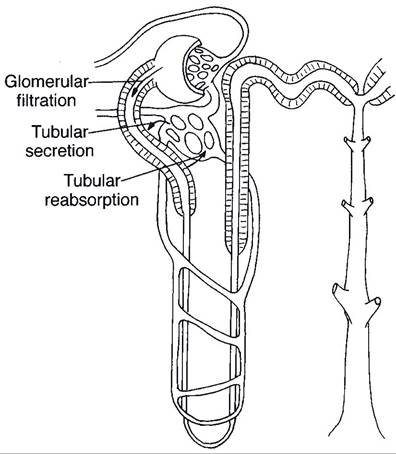
Figure 23-7. Urine formation. The glomerular filtrate enters the tubules, where some substances are removed by selective tubular reabsorption and others are added by selective tubular secretion. Reabsorption and secretion occur throughout the nephron and collecting ducts. (Reprinted with permission of Wiley-Blackwell from Reece W.O. Physiology of Domestic Animals. 2nd ed. Baltimore: Williams & Wilkins, 1997.)
glomeruli, a large quantity of filtrate is formed and enters the urinary space of Bowman’s capsule. From here the filtrate flows through the tubules and collecting ducts, where tubular reabsorption and tubular secretion alter its volume and composition. Tubular reabsorption is the removal of substances from the tubular fluid by the tubular cells; these substances are usually returned to the blood in the peritubular capillaries. Tubular secretion is the addition of substances to the tubular fluid by tubule cells. The secreted substances are produced in the tubule cells (e.g., hydrogen ion and ammonia) or taken up by the tubule cells from the blood in the peritubular capillaries (e.g., pharmaceuticals).
The renal microcirculation is unique in that glomerular capillaries are between two arteriolar vessels rather than between an arteriole and a venule. Afferent arterioles lead into glomer-
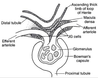
Figure 23-8. The juxtaglomerular (JG) apparatus. Extraglomerular mesangial cells are found at the base of the glomerulus between the macula densa and the arterioles.
(Reprinted with permission of Wiley-Blackwell from Reece W.O. Physiology of Domestic Animals. 2nd ed. Baltimore: Williams & Wilkins, 1997.)uli, and efferent arterioles leave glomeruli (Fig. 23-4). Efferent arterioles from most glomeruli lead into capillary networks that surround tubules in the cortex (peritubular capillaries). Efferents from glomeruli deep in the cortex next to the medulla contribute blood to vessels that extend into the medulla. These vessels (vasa rectae) consist of straight descending branches (descending vasa rectae) that empty into medullary capillaries, which are drained by straight ascending vessels (ascending vasa rectae) (Fig. 23-4).
Near glomeruli, the walls of afferent arterioles contain specialized cells termed juxtaglomerular (JG) or granular cells (Fig. 23-8). secretory granules in these cells contain the enzyme renin. Renin is a component of the renin-angiotensin-aldosterone system (see Chapter 18), which is involved in the regulation of blood volume and blood pressure. The JG cells are part of a functional grouping of closely related structures, the juxtaglomerular apparatus. The juxtaglomerular apparatus consists of the JG cells, the macula densa, and extraglomerular mesangial cells (Fig. 23-8). The macula densa is a specific region of the wall of the distal tubule where the cellular nuclei appear to be bunched closely together. The segment of the distal tubule found here is part of the same nephron associated with the afferent arterioles (Fig. 23-7). The extraglomerular mesangial cells are between the macula densa and its associated JG cells.