Oxytocin and Vasopressin Are Synthesized in Cell BodiesWithin the Hypothalamus and Are Carried by Axon Flow to the Posterior Lobe, Where They Are Released
As indicated, the two important hormones produced by the neurohypophysis are vasopressin and oxytocin. Although it was previously thought that the two hormones were produced in separate nuclei* evidence now indicates that both hormones are produced in both the supraoptic and the paraventricular nucleus.
The cell bodies that synthesize the hormones are large and thus are called magnocellular nuclei. The synthesis of vasopressin and oxytocin, as described previously for protein and peptide hormones, involves first the production of a preprohormone, prepropressophysin for vasopressin and prepro-oxyphysin for oxytocin, at the level of the cell body within the hypothalamus (Figure 33-12). The “pre” portion of the molecule is cleaved before the molecules are packaged into granules. During passage of the granules down the axon, the prohormone is cleaved to produce either oxytocin or vasopressin; the remaining peptide fragments are called neuro- physin I or neurophysin II, respectively. Neurophysin I, which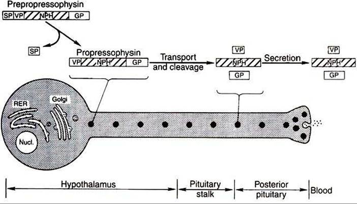
FIGURE 33-12 Diagramofa vasopressin-secreting neuron illustrating the Subcellular components involved in synthesis and secretion. This process begins with the synthesis of prepropressophysin, which consists of (1) a signal peptide (SP), (2) vasopressin (VP), (3) neurophysin (NPH)1 and (4) a glycoprotein ^GPJ.The production and release of oxytocin is identical except that no glycoprotein is involved. RER, Rough endoplasmic reticulum; Nucl., nucleus. (From Hedge GA, Colby HD, Goodman RL: Clinical endocrine physiology, Philadelphia, 1987, Saunders.)
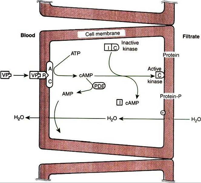
FIGURE 33-13 Antidiuretic mechanism of action of vasopressin (VP) on cells of the distal tubule and collecting ducts.
AC, Adenyl cyclase; AMP, adenosine monophosphate; ATP, adenosine triphosphate; cAMP, cyclic AMP;
/and C, inhibitory and catalytic subunits of the kinase, respectively; PDE, phosphodiesterase; R, receptor. (From Hedge GA, Colby HD, Goodman RL: Clinical endocrine physiology, Philadelphia, 1987, Saunders.)
is released into the vascular system along with oxytocin, has been quantified as an alternative means of following the release of oxytocin. At present, the physiological function of the neurophysins is not known.
The release of the posterior lobe peptide hormones is initiated in the hypothalamus as a result of depolarization of the cell body because of stimulation by neural afferents. The action potential generated extends down the axon to the nerve terminal, where the secretory granules containing the hormone are stored. The depolarization of the nerve cell membrane allows the influx of calcium ions, which initiates the release of hormone th rough the process of exocytosis.