Peripheral Nerves
The central nervous system, the brain and spinal cord, is in two-way communication with virtually all body tissues by means of a system of branching peripheral nerves. These are composed of afferent (sensory) fibers, which convey information to the central nervous system from peripheral receptors, and efferent (motor) fibers, which convey instructions from the central nervous system to peripheral effector organs.
The peripheral nerves comprise the 12 pairs of cranial nerves and the considerably larger number of pairs of spinal nerves whose total varies with the vertebral formula. The dog has 8 cervical, 13 thoracic, 7 lumbar, 3 sacral, and about 5 caudal pairs. The present discussion is restricted to the rather uniform spinal nerves; the cranial nerves differ from these and from one another in many respects that are considered later (p. 301).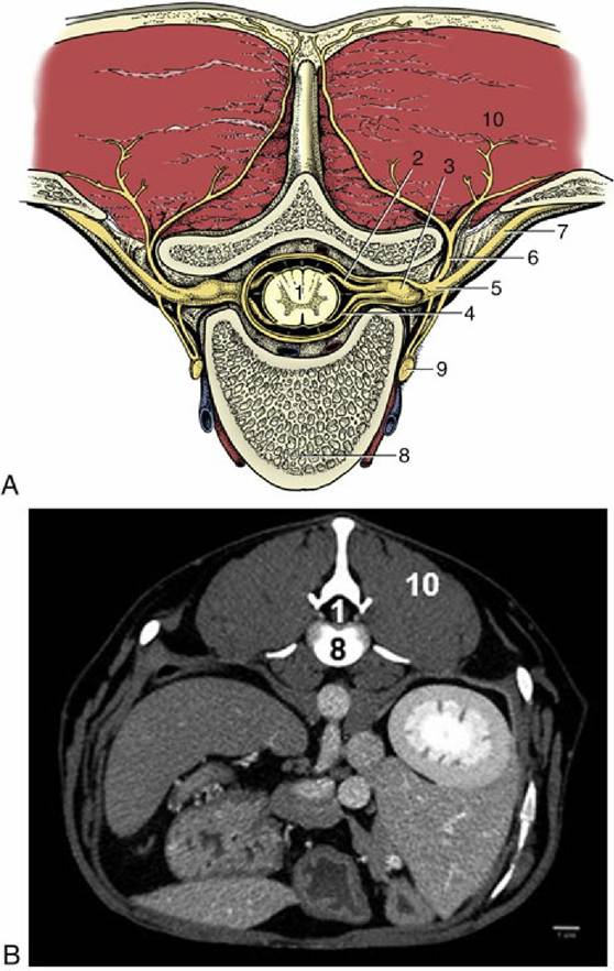
FIG. 1.36 Schematic (A) transection of the vertebral column to show the formation of a spinal nerve. B shows a CT of the abdomen of a female dog. 1, Spinal cord; 2, dorsal root; 3, spinal ganglion; 4, ventral root; 5, spinal nerve; 6, dorsal branch of spinal nerve; 7, ventral branch of spinal nerve; 8, body of vertebra; 9, sympathetic trunk; 10, epaxial muscles.
The orderly origin of the spinal nerves reveals the segmentation of the spinal cord. Each nerve is formed by the union of two roots (Fig. 1.36). The dorsal root is almost exclusively composed of afferent fibers whose cell bodies are clumped together to form a visible swelling, the spinal (dorsal root) ganglion. The central processes enter the cord along a dorsolateral furrow. The peripheral processes extend from the wide variety of exteroceptive, proprioceptive, and enteroceptive endings that respond to external stimuli, changes within the muscles and other locomotor organs, and changes in the internal organs, respectively.
The ventral root is exclusively composed of efferent fibers emanating from motor neurons within the ventral horn of gray matter and leaving the cord along a ventrolateral strip; they are in passage to the effector organs—muscles and glands.The dorsal and ventral roots join peripheral to the dorsal root ganglion to form the mixed spinal nerve (Fig. 1.36/5), which leaves the vertebral canal through the appropriate intervertebral foramen. In the cervical region, each nerve emerges cranial to the vertebra of the same numerical designation as the nerve, except the eighth, which emerges between the last cervical and first thoracic vertebrae. In other regions, each nerve emerges caudal to the vertebra of the same numerical designation.
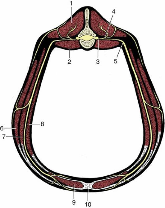
FIG. 1.37 The distribution of a (lumbar) spinal nerve. 1, Epaxial muscles; 2, sublumbar muscles; 3, spinal nerve; 4, dorsal branch of spinal nerve; 5, ventral branch of spinal nerve; 6, 7, external and internal abdominal oblique muscles; 8, transversus abdominis muscle; 9, rectus abdominis muscle; 10, linea alba.
The mixed trunk formed by the union of dorsal and ventral roots divides almost at once into dorsal and ventral branches (rami). The dorsal branch is distributed to dorsal structures: epaxial muscles of the trunk (broadly, those that lie dorsal to the line of transverse processes) and the skin over the back (Fig. 1.37). The much larger ventral branch is distributed to hypaxial muscles of the trunk (broadly, those ventral to the transverse processes), the muscles of the limbs (with a few exceptions), and the remaining part of the skin, including that of the limbs. Both dorsal and ventral branches have connections with their neighbors that form continuous dorsal and ventral plexuses. These plexuses are generally neither obvious nor important, except for enlarged portions of the ventral plexus opposite the origins of the limbs.
These, the brachial and lumbosacral plexuses, give rise to the nerves that are distributed to forelimb and hindlimb structures, respectively.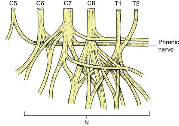
FIG. 1.38 The brachial plexus. The ventral divisions of the spinal nerves (C6 through T2) contributing to the plexus are at the top of the schema, the peripheral branches (N) supplying the forelimb at the bottom. Contributions from C5, C6, and C7 form the phrenic nerve.
The brachial plexus (Fig. 1.38) is usually formed by contributions from the last three cervical and the first two thoracic nerves and the lumbosacral plexus by contributions from the last few lumbar and the first two sacral nerves. The limb plexuses allow for regrouping and reassociation of the constituent nerve fibers, and the nerve trunks that emerge distally are each composed of fibers derived from two or three spinal segments; thus the median nerve is composed of fibers from spinal nerves C8 and T1, the femoral nerve of fibers from L4 to L6.
The courses of the major peripheral nerve trunks must be known to avoid placing the nerves at unnecessary risk during surgery. Their central connections are important in two contexts. First, local anesthetic solutions injected near selected spinal nerves have predictable effects in paralyzing muscles and in depriving skin areas of sensation. Conversely, paralysis of particular muscles or absence or alteration of the sensibility of specific skin areas may point to the precise location of a central lesion.
So far, reference to nerve fibers concerned with the innervation of blood vessels, glands, and internal organs has been avoided. These structures are supplied by the autonomic division of the nervous system, which is described in Chapter 8. For the present, it is sufficient to state that although autonomic fibers are not present in the roots of every spinal nerve, arrangements exist that ensure that each peripheral nerve receives its necessary quota.
Comprehension Check
Is a knowledge of anatomy critical for a person to become a competent veterinary medical professional? If so, list arguments to support your position.
>------------------------- <
* There is a separate but similar vocabulary (Nomina Anatomica Avium) that is concerned with the anatomy of birds.
* Among domestic mammals, horses and cattle have synovial fossae. Although not quite constant, synovial fossae appear in the majority of animals and are always bilateral in the limbs. They appear as early as 10 days after birth in foals. In the horse, opposing synovial fossae are found at the shoulder, elbow, carpal, tarsocrural, and talocalcaneal joints. A single fossa is present in the fetlock joints (of both forelimbs and hindlimbs), on the acetabulum, and on the atlantal surface of the atlanto- axial joint. In cattle, more or less distinct synovial fossae may be present in all limb joints, other than the shoulder and hip. They also may be present at the atlantooccipital and atlanto- axial joints