Pharynx
The pharynx (pl. pharynges) is the common passage for food and air, is caudal to oral and nasal cavities, and is lined by mucous membrane and surrounded by muscles. The pharynx can be arbitrarily divided into nasal (nasopharynx), oral (oropharynx), and laryngeal (laryn- gopharynx) portions, so named for their association with these regions.
The muscles of the walls of the pharynx are responsible for the orderly directing of air, food, and liquids in such a way that air from the nasal cavity is directed into the ventral larynx and food and liquids are directed into the dorsal esophagus. Thus, the paths of air and swallowed substances must cross in the pharynx; pharyngeal dysfunction can have severe consequences for the airway, which must be protected from foodstuffs (Fig. 20-6).
The pharyngeal recess in the horse is a median niche at the caudodorsal angle of the nasopharynx (Fig. 20-7). The pig has a pharyngeal diverticulum that opens into the dorsal wall of the pharynx near the beginning of the esophagus.
Care must be exercised not to enter this diverticulum while passing a stomach tube or giving medications via a balling gun.
Tonsils
Tonsils are more or less circumscribed aggregations of lymphatic nodules found in association
Figure 20-6. Relationship of pharynx and mouth to larynx and esophagus during (A) normal respiration and (B) swallowing.
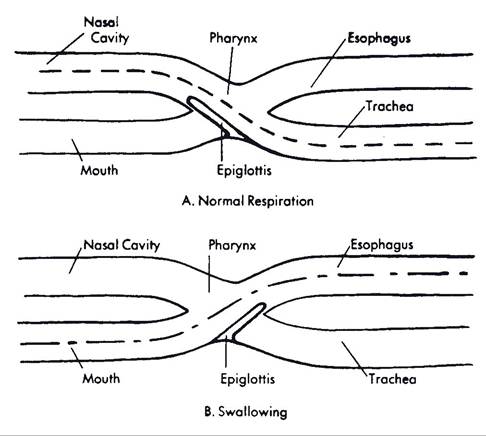
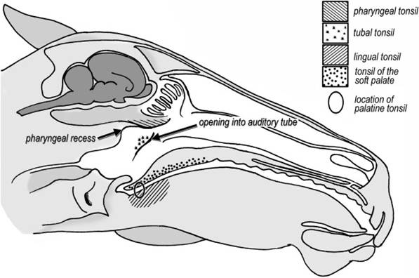
Figure 20-7. Sagittal section of the equine head, showing locations of tonsils.
with the mucous membranes of the mouth and pharyngeal region (Fig. 20-7). The histologic organization of tonsils is described in Chapter 16.
In humans, each of two obvious palatine tonsils lies in a pocket on the lateral wall of the pharynx ventral to the soft palate and adjacent to the base of the tongue (these are readily seen with a mirror as elongate nodules on each side of your throat, unless, of course, they have been removed).
in the horse, ox, and sheep, these palatine tonsils are in about the same relative position, within the submucosa, and completely covered by mucous membrane. in other words, the tonsils do not project into the pharynx at all in these animals. The ruminant palatine tonsil features a deep cleft, the tonsillar crypt, which increases the surface area through which the lymphatic tissue may come in contact with antigens (Fig. 20-4). In the pig the palatine tonsils lie in the substance of the soft palate.The lingual tonsils consist of accumulations of lymphatic nodules in the base of the tongue. These tonsils are most prominent in the horse, ox, and pig.
The pharyngeal tonsil is an accumulation of lymphoid tissue in the submucosa of the dorsal pharyngeal wall of all domestic animals. Enlargement of the pharyngeal tonsil of humans creates the condition commonly called adenoids.
The openings of the auditory tubes (see Chapter 11) in the nasopharynx feature aggregations of lymphatic nodules, the tubal tonsils.