Mouth
The mouth is used primarily for holding, grinding, and mixing food with saliva but may also be used to manipulate the environment (through grasping of objects) and as a defensive and offensive weapon.
The entrance into the mouth is defined by the lips (labia), the appearance and mobility of which vary among species. The external parts of the lips are covered by typical haired skin, which changes to mucous membrane at the mucocutaneous junction of the labial margins. The upper lip of small ruminants is deeply grooved with a midline philtrum. Lips are densely innervated by sensory fibers, making them very sensitive tactile organs. The lips of sheep, goats, and horses are soft and flexible and aid in picking up food, whereas those of cattle and hogs are stiffer and less mobile.
The small space between the teeth and lips is the oral vestibule. The oral cavity proper lies deep to the teeth and is occupied primarily by the tongue. The oral cavity ends at a narrowing (the isthmus of the fauces) near the base of the tongue, where the digestive tract continues as the pharynx.
The dorsal wall of the oral cavity comprises the hard palate rostrally and the soft palate caudally. The hard palate is formed by horizontal elements of the incisive, maxillary, and palatine bones, and its thick mucous membrane covering is characterized by prominent transverse folds called palatine rugae. The soft palate is a musculomucosal sheet that extends toward the base of the epiglottis (see Chapter 19). The equine soft palate is exceptionally long, and the horse is uniquely unable to actively lift the soft palate so as to permit passage of air from the oral cavity to the larynx. For this reason, horses are obligate nose breathers, breathing through the mouth only when the soft palate is displaced dorsad from its normal position ventral to the epiglottis.
Teeth
Teeth are arranged in two dental arcades, one associated with the mandible and one with the incisive and maxillary bones. Farm animals typically have a gap in each arch between the front teeth (incisors) and the cheek teeth (see Fig. 4-4); such a physiologic gap is a diastema. The bit of the bridle lies in the horse’s diastema.
Mammals typically exhibit heterodonty. That is, they have various types of teeth that are specialized for different aspects of prehension and mastication. All domestic animals also are diphyodont. This means they develop a set of deciduous teeth (also called baby teeth or milk teeth) that fall out and are replaced with permanent teeth. As growing teeth emerge from the gums, they are said to erupt. When their occlusal (grinding) surfaces meet those of the teeth in the opposing arcade, they are said to have come into wear. Eruption times of teeth are consistent enough to permit accurate aging of young animals by observing their dentition (see Tables 20-1 and 20-2). The age of animals with a full set of permanent teeth can be estimated through examination of the wear patterns of the occlusal surfaces.
A tooth is anchored by its root in a socket of bone called an alveolus. A connective tissue, the periodontium (also called periodontal membrane), firmly attaches the root to the surrounding bone in a specialized joint, the gomphosis (see Chapter 6). The crown is the part
| Table 20-1. Formulas and Eruption of Deciduous Teeth | |||||
| Deciduous Teeth | Horse | Ox | Sheep | Pig | |
| Formulas* | 30 | 3 | 003 | 003 | 314 |
| 3 0 | 3 | 4 0 3 | 4 0 3 | 3 1 4 | |
| Eruption of Incisors | |||||
| DI 1 | birth to | 1 week | birth to 2 wk | birth to 1 wk | 2-4 wk |
| DI 2 | 4-6 wks | birth to 2 wk | birth to 1 wk | 1.5-3 mo | |
| DI 3 | 6-9 mo | birth to 2 wk | birth to 1 wk | birth or before | |
| DI 4 | birth to 2 wk | birth to 1 wk | |||
| Eruption of Canines | before birth | ||||
| Eruption of Premolars DP 1 | birth to few days | usually before birth | 3.5-6.5 mo | ||
| DP 2 | birth to | 2 wk | birth to few days | usually before birth | 7-10 wk |
| DP 3 | birth to | 2 wk | birth to few days | usually before birth | 1-3 wk (upper) 1-5 wk (lower) |
| DP 4 | birth to | 2 wk | 1- 4 wk (upper) 2- 7 wk (lower) | ||
*The deciduous formula is shown for one side of the mouth only, with upper arcade represented above and lower arcade below.
The first number is the incisors; the second, canines; and the third is premolars.
Table 20-2. Formulas and Eruption of Permanent Teeth
| Permanent Teeth | Horse | Ox | Sheep | Pig | ||||||||
| Formulas* | 3 | 1 | 3-4 | 3 | 0 0 | 3 | 3 | 0 0 | 3 | 3 | 3 1 4 | 3 |
| 3 | 1 | 3 | bgcolor=white>34 0 | 3 | 3 | 4 0 | 3 | 3 | 3 1 4 | 3 | ||
| Eruption of Incisors | ||||||||||||
| I 1 | 2.5 | yr | 1.5-2 yr | 1-1.5 yr | 1 yr | |||||||
| I 2 | 3.5 | yr | 2-2.5 yr | 1.5-2 yr | 16-20 mo | |||||||
| I 3 | 4.5 | yr | 3 yr | 1.5-3 yr | 8-10 mo | |||||||
| I 4 | 3.5-4 yr | 3.5-4 yr | ||||||||||
Eruption of Canines
4-5 yr 9-10 mo
Eruption of Premolars
| P 1 | 5-6 mo | 2-2.5 yr | 1.5-2 yr | 12-15 mo |
| P 2 | 2.5 yr | 1.5-2.5 yr | 1.5-2 yr | 12-15 mo |
| P 3 | 3 yr | 2.5-3 yr | 1.5-2 yr | 12-15 mo |
| P 4 | 4 yr | 12-15 mo | ||
| Eruption of Molars | ||||
| M 1 | 9-12 mo | 5-6 mo | 3-5 mo | 4-6 mo |
| M 2 | 2 yr | 1-1.5 yr | 9-12 mo. | 8-12 mo |
| M 3 | 3.5-4 yr | 2-2.5 yr | 1.5-2 yr | 18-20 mo |
*The permanent formula is shown for one side of the mouth only, with upper arcade represented above and lower arcade below. The first number is the incisors; the second, canines; the third is premolars; and the fourth is the molars.
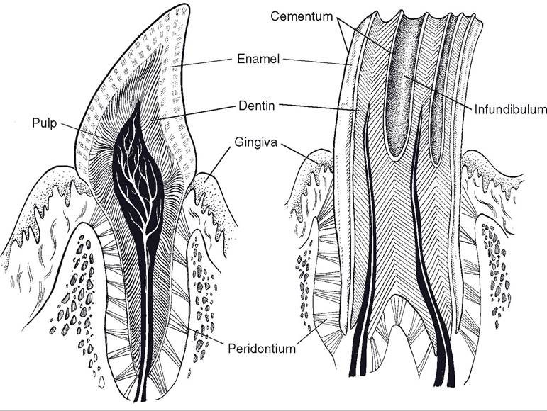
Figure 20-2.
Anatomy of typical brachyodont (left) and hypsodont (right) teeth.of the tooth visible above the mucous membrane of the gum. Some teeth have a short crown separated from the root by a distinct neck. These teeth, of which ruminant incisors are an example, are described as brachyodont (from the Greek word brachy, short). In contrast, equine incisors and cheek teeth have a tall, straight crown with no discernible neck. These teeth are described as hypsodont (Greek hypsi, high) (Fig. 20-2).
Most of the tooth’s substance is made up of a mineralized substance called dentin (sometimes spelled dentine), with a dental cavity at its center. The connective tissues, nerves, and blood vessels of the tooth reside in this cavity and constitute the dental pulp. It is dentin, incidentally, that constitutes the “ivory” of elephant tusks.
Superficial to the dentin is a layer of enamel, a white layer consisting of inorganic crystals. Enamel is the hardest substance in the body. It also is irreplaceable, as the cells that generate it (ameloblasts) are lost following formation of the tooth, the only exception amongst farm animals being the tusks (canine teeth) of swine. only the crowns of brachyodont teeth are covered with enamel, whereas nearly the entire hypsodont tooth has a layer of enamel, with only a small, short region of the tooth’s root lacking this layer. The enamel of hypsodont teeth is thrown into prominent folds on the grinding surfaces of these teeth, where they form characteristic crests (cristae enameli) and cups (infundibula).
Cementum is a thin, bonelike layer on the surface of the tooth. It covers only the root of the brachyodont tooth, but it extends from the root to cover the crown of the hypsodont tooth.
The front teeth are called incisors, and in dental formulas, they are designated by the letter I. The incisors are numbered starting from the midline. The first pair of incisors is called I1, or centrals; the next pair I2, or first intermediates; next I3, or second intermediates; and the last and most lateral pair of incisors is called I4, or corners.
In the nonruminants, only one pair of intermediate incisors is found. Ruminants lack incisors in the upper dental arcade. Instead, the mucous membrane in this region is modified into a dense, keratinized dental pad, against which the lower incisors abut when the jaws are closed. Permanent incisors are preceded by a like number of deciduous teeth.Canine teeth (abbreviated C) are also called eyeteeth, bridle teeth, tusks, and tushes. Ruminants lack canine teeth, and although they can be well developed in stallions, they are small or absent in mares and geldings. The canine teeth of pigs are large, especially in boars, and in this species they are usually called tusks. Porcine tusks are described as open rooted, meaning that they continue to grow throughout life. The lower tusk is generally much larger than its partner in the upper arcade. in pigs, the permanent canines are preceded by analogous deciduous teeth; in the horse, the deciduous canines are often absent or so small that their crowns do not erupt.
Cheek teeth comprise premolars (P) and molars (M), which in herbivores are morphologically similar. only premolars are preceded by deciduous teeth; the molars have no precursors. in the pig, the molars are larger than the premolars and have a flatter occlusal surface, in keeping with their grinding function.
The first premolar of horses is often absent, and when present, it is almost always seen only in the upper (maxillary) arcade. Unlike the other equine cheek teeth, the first upper premolar is very small and does not come into wear. This tooth is called the wolf tooth, and some horse owners prefer to have it removed because of the perception that it may make unwanted contact with the bit.
In the young horse, only a small part of each cheek tooth is visible, as most of the crown lies developed but unerupted beneath the gum (Fig. 20-3). Throughout the horse’s life, these teeth continue to erupt, maintaining their intraoral height even as they are
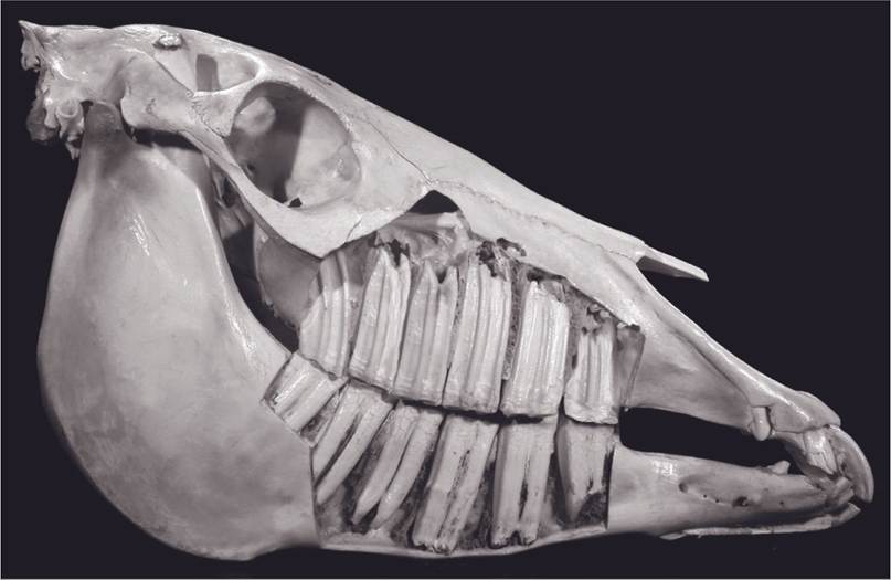
Figure 20-3.
Hypsodont teeth. The cheek teeth of the young horse are fully formed but largely within the bones of the face and mandible. The teeth slowly erupt over the life of the horse as the crowns are worn by chewing of coarse feedstuffs.worn down by the coarse forage the horse eats. Equine cheek teeth are not open rooted; no new dental tissues are created after the tooth is initially formed. They are instead described as slowly erupting. Because of this phenomenon, horses are prone to develop sharp, elongated ridges of enamel on their cheek teeth. These are variously called points and hooks, and they may cause the horse considerable pain if they cut the soft cheeks, tongue, and gums. For this reason, it is considered good husbandry to file down the points and hooks periodically, a procedure called floating the teeth that is done with a special rasp called a fl oat.
Deciduous and permanent dental formulas for domestic farm animals are shown in Tables 20-1 and 20-2.
Tongue
The tongue consists of a mass of muscle covered by mucous membrane. it is divided into a free apex at the rostral end, a meaty body, and a caudal root adjacent to the pharynx. The entire tongue is mobile through its muscular attachments to the hyoid apparatus and mandible. The muscles of the tongue (intrinsic muscles) have fibers oriented in longitudinal, perpendicular, and transverse directions, permitting the tongue a wide range of movements. This is particularly evident in the ox, which uses its tongue as a prehensile organ (Fig. 20-4).
The tongue is covered with thick keratinized stratified squamous epithelium. The surface is characterized by a large number of projections, the papillae, which are particularly well developed on the dorsal surface (Fig. 20-5). Filiform, fungiform, and vallate papillae are found in all domestic animals, and foliate papillae are present in the horse, pig, and dog, but not in ruminants. Ruminants additionally have large conical papillae. The filiform and conical papillae do not bear taste buds (cells specialized for gustation; see Chapter 11), but all other types of papillae do. Taste buds may also be found on the epiglottis, larynx, pharynx, and soft palate.
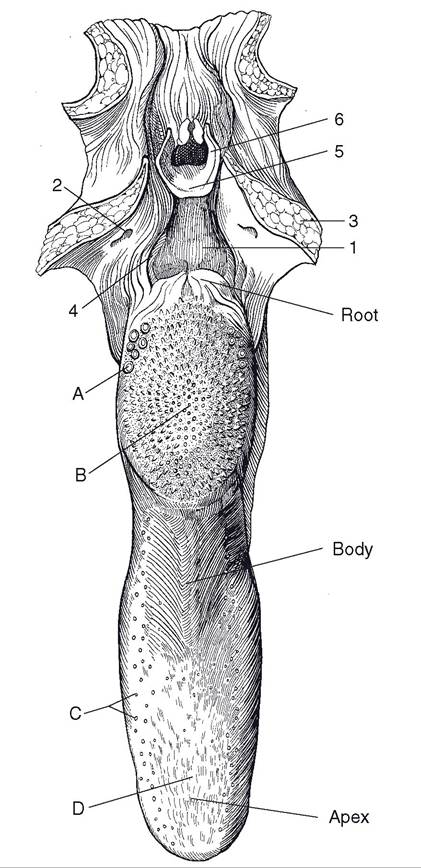
Figure 20-4. Tongue of the ox, dorsal view. The pharynx and soft palate are transected and reflected laterad. A, vallate papillae; B, torus linguae with conical papillae; C, fungiform papillae; D, filiform papillae; 1, oropharynx; 2, tonsillar crypt; 3, cut surface of soft palate; 4, palatopharyngeal arch; 5, epiglottis; 6, opening into larynx.
The filiform papillae look somewhat hairlike. in the ox, they consist of a connective tissue core covered by a highly cornified epithelial layer. These papillae are shorter and softer
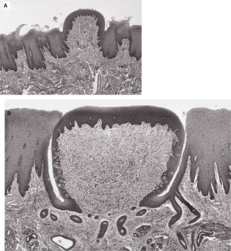
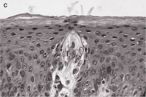
Figure 20-5. A) Fungiform and filiform papillae of the caprine tongue. The keratinized filiform papillae have no taste buds. B) Vallate papilla of the caprine tongue. Note the deep groove adjacent to the papilla; taste buds occur in the mucosa of the papilla of this groove. C) Higher magnification of a taste bud from an equine fungiform papilla. (Reprinted with permission of Wiley-Blackwell from Bacha, Jr. W.J. and Bacha L.M. Color Atlas of Veterinary Histology. Baltimore: Lippincott Williams & Wilkins, 2000.)
in the horse than in other domestic animals, giving the tongue of the horse its velvety feel. Interspersed amongst the filiform papillae are fungiform papillae, so called because of their resemblance to tiny mushrooms.
Foliate papillae resemble the foliage or leaves of plants. They are found in the horse and pig (and only rarely in cattle) on the lateral margin adjacent to where the root of the tongue is connected to the soft palate by a mucous membrane fold, the palatoglossal arch.
Vallate papillae are large, circular projections surrounded by a deep groove. These papillae are arranged in a v shape on the caudal part of the tongue and demarcate the morphologic division between the body and the root of the tongue.
The body of the ruminant tongue has a prominent dorsal bulge, the torus lingua, which is thickly covered with prominent conical papillae. Similar cornified projections cover the inside of the lips and cheeks.