PHYSICOCHEMICAL PROPERTIES OF SOLUTIONS
1. How does facilitated diffusion differ from simple diffusion?
2. What parts of a cell membrane (protein or lipids) account for the diffusion of water-soluble substances? What parts are considered to be the pores?
3.
How does active transport differ from facilitated diffusion?4. Define osmosis.
5. Define a semipermeable membrane.
6. Define osmotic pressure and how it is determined.
7. How does a selectively permeable membrane differ from a semipermeable one?
8. As related to tone, how does effective osmotic pressure for a solution differ from a measured osmotic pressure?
9. What is the difference between hemoglobinemia and hemoglobinuria?
An understanding of the physicochemical properties of solutions can be considered to be just as important to the study of physiology as knowledge of anatomy. These properties will be encountered when studying most of the systems that follow. Further, successful fluid replacement and correction of deficiencies in live animals require an understanding of solution composition and effects of their administration. When H.F. Weisberg approached this topic, he seemed to have a feel for its basic importance when he quoted the following biblical verse (Proverbs 4:7): “Wisdom is the principal thing; therefore get wisdom: and with all thy getting get understanding.” It is in this context that the basic principles of physicochemical properties of solutions are presented, with emphasis on understanding.
Diffusion
Simple diffusion refers to the random movement of molecules, ions, and suspended colloid particles under the influence of Brownian (thermal) motion. Brownian motion is observed when light shines through air and dust particles are seen to move in a random motion. The movement of the dust particles is caused by their bombardment by air molecules. This same random motion occurs between air and dust or between two different metals placed side by side.
Over time, the two metals will fuse with each. This is still simple diffusion. If a concentration gradient (differential) exists, molecules, ions, and colloidal particles tend to move from the area of their higher concentration to the area of their lower concentration, “downhill.” The movement is specific to each substance (i.e., Na+ will diffuse from the area of its higher concentration to the area of its lower concentration regardless of the presence and concentrations of other substances). If the molecules and ions are dispersed equally, the random motion continues but does not accomplish net movement or flow; this represents a state of equilibrium. Energy is not required for simple diffusion.Barriers to diffusion are generally the membranes of cells. These consist of a lipid bilayer, which is a thin film of lipid only two molecules thick through which fat-soluble substances (especially carbon dioxide and oxygen) can readily diffuse (Figure 2-1). There might be facilitated diffusion for other substances, in which a carrier is required (Figure 2-2). However, facilitated diffusion for any substance still occurs from the area of its higher concentration to that of its lower concentration and, as in simple diffusion, energy is not required. Because cell membranes are predominantly lipid, they are relatively hydrophobic (water repelling) and the diffusion of water through the lipid bilayer proceeds with difficulty; however, water can diffuse through protein channels. Protein channels (Figure 2-1) consist of large protein molecules interspersed in the lipid film; they provide structural pathways (pores) not only for water but also for water-soluble substances. Some substances may be excluded from diffusion through the pores because of their large size; conversely, diffusion may be facilitated because of other factors, such as a substance’s relatively smaller size, its electrical charge (e.g., negative pore charge assists Na+ diffusion), or the protein channel’s specificity (e.g., specific ion channels).
Other protein channels act as carrier proteins for the transport of substances in a direction opposite to their natural diffusion pathway. This is known as active transport. Whereas the transport of glucose into most cells of the body is accomplished by facilitated diffusion, exceptions exist in the lumens of the kidney tubules and intestines, where active transport is involved. In these locations, glucose is continually transported into the blood, where the concentration is high, from the lumen where its concentration may be minute. Loss of glucose from the body is prevented in these locations because of active transport. Active transport requires not only a carrier but also energy.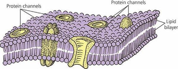
■ FIGURE 2-1 Structure of a cell membrane. The lipid bilayer is represented by a thin film of lipid that is two molecules thick. The protein channels (pores) may be composed of a single protein or a cluster of proteins. The channels may have specificity for certain substances, or they may be restrictive because of size. Virtually all water diffuses through the protein channels.
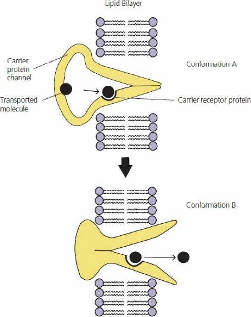
■ FIGURE 2-2 A postulated mechanism for facilitated diffusion. A. The transported molecule enters the protein channel and binds with the receptor at the binding site. B. Subsequent to binding, the protein channel undergoes a conformational change to open the channel on the opposite side and the transported molecule is released, causing return of the protein channel to its original conformation.
Osmosis and Osmotic Pressure
The most abundant substance in the body that undergoes diffusion is water. Diffusion of water occurs throughout the body relatively easily. The amount diffusing into cells is usually balanced by an equal amount diffusing out. Osmosis is the process of diffusion of water through a Semipermeable membrane from a solution of higher water concentration to a solution of lower water concentration.
A semipermeable membrane is one that is permeable (permits passage) to water but not to solutes. When comparing water concentrations of solutions, it is implied that the solution with the highest water concentration has the lowest solute concentration. A situation in which osmosis could occur is illustrated in Figure 2-3 where two different concentrations of water are separated by a semipermeable membrane. Net diffusion has occurred from the compartment with the highest water concentration, compartment 1, to the one with the lowest water concentration, compartment 2.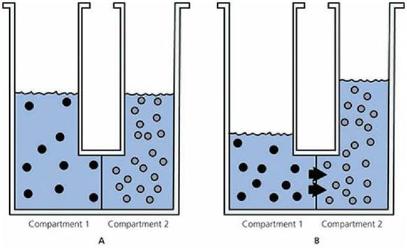
■ FIGURE 2-3 Osmosis. A. Before osmosis. Equal volumes of aqueous solutions (solutes represented by black circles and open circles) are placed in compartments that are separated by a membrane permeable to water but not to the solutes (semipermeable membrane). The aqueous solution in compartment 1 has the highest concentration of water (lowest concentration of solute). B. During osmosis. Osmosis (diffusion of water) occurs from compartment 1 to compartment 2 (highest water concentration to lowest water concentration) and the water level rises in compartment 2.
The quantitative measure of the tendency for water to osmose is osmotic pressure. In the above example, this is the downward pressure that would have to be applied to the compartment with the lowest concentration of water (compartment 2) to prevent net diffusion of water from the compartment with the highest water concentration (compartment 1). In the animal body, osmotic pressure is a potential pressure because osmosis is not prevented when water imbalances exist. The number of particles in a solution (i.e., ions, molecules) determines its osmotic pressure; the greater the number of particles, the higher the osmotic pressure. For two aqueous solutions of NaCl separated by a membrane that permits diffusion of water but not NaCl, the highest osmotic pressure is measured for the solution with the highest concentration of NaCl (lowest concentration of water).
Water will diffuse to the area of greatest osmotic pressure.Osmolar concentrations are used to express the osmotic strength of solutions (e.g., urine, plasma, NaCl). One mole of an undissociated (not ionized) substance is equal to 1 osmole (osmol). If a substance dissociates into two ions (NaCl → Na+ and Cl-), 0.5 mol of the substance equals 1 osmol. The number of particles, not the mass of the solute, determines osmotic pressure. One liter of a solution that contains 300 milliosmol of 0.3 mol/L of glucose (which does not dissociate) exerts the same osmotic pressure as one that contains 300 mOsm of 0.15 mol/L of NaCl. Similarly, the osmolality of a urine sample (many substances, both ionized and undissociated) measured as 300 mosmol exerts the same osmotic pressure as the previous solutions of glucose and NaCl.
A comparison of osmotic pressure for several solutions is shown in Table 2-1. Values were determined using an osmometer and are given in osmolality (mosmol/kg H2O). An osmometer is an instrument for measuring osmolality by freezing point depression or vapor pressure lowering techniques (colligative properties). Values obtained are representative of diffusion through semipermeable membranes. Note that bovine urine has an osmotic pressure 3.3 times greater than that of bovine plasma (water concentration lower, solute concentration greater than bovine plasma). Canine urine has an osmotic pressure 6.1 times greater than that of canine plasma. Urine is formed from plasma and canines have a greater potential for concentrating urine than bovines.
TABLE 2-1 OSMOLALITY OF SEVERAL SOLUTIONS AS DETERMINED BY VAPOR PRESSURE LOWERING OSMOMETRYa
| SOLUTION IDENTIFICATION | OSMOLALITY.(mOsm/kg H2O) |
| Bovine plasma | .302 |
| Bovine urine | 1,031 |
| Bovine milk (skim) | .272 |
| Canine plasma | .312 |
| Canine urine | 1,904 |
| Tap H2O | .58 |
| aValues obtained from student laboratory exercises. | |
Tone of Solutions
The membranes of the body vary in their permeabilities and allow certain solutes (as well as water) to diffuse through them.
They are selectively permeable membranes. The measured osmotic pressure for a solution containing solutes that could diffuse through membranes would then not be an index for its tendency to cause osmosis. Instead, the tone of a solution is defined, which is the effective osmotic pressure. Only those particles (molecules, ions) for which the membrane is not permeable contribute to the tone. The principles of osmosis continue to prevail, except that now water diffuses to the greatest effective osmotic pressure. Figure 2-4 illustrates the tone of solutions. Two solutions of equal volumes and particle numbers are shown to be separated by a membrane that permits the passage of water and the particles in compartment 2. Each solution has the same measured osmotic pressure (same concentration of particles). Because compartment 1 has particles that cannot diffuse through the membrane, these particles are the ones that contribute to an effective osmotic pressure and, because the solution in compartment 2 has no effective osmotic pressure (because particles are diffusible), water diffuses to the greatest effective osmotic pressure, or from compartment 2 to compartment 1. In this example the net diffusion of water stops when the pressure resulting from the weight of the solution in compartment 2 opposes the diffusion resulting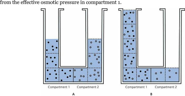
■ FIGURE 2-4 Hypothetical example of the tone of solutions. A. Before osmosis. Two aqueous solutions (solutes represented by black circles and open circles) of equal osmotic pressure are separated by a membrane permeable to water and open circle solutes (selectively permeable membrane). B. During osmosis. Effective osmotic pressure is exerted only by black circle solutes, and water diffuses from compartment 2 to compartment 1. At equilibrium, an open circle solute has a new, lower concentration that is equal throughout compartments 1 and 2. (Dashed lines represent divisions of equal volume.)
From a practical standpoint, the tone of a solution that can be infused into the blood of animals is usually compared with the solution inside red blood cells (erythrocytes). The solution of erythrocytes is in osmotic equilibrium with plasma (the fluid part of blood). An infused solution is hypotonic if it has a lower effective osmotic pressure than the solution of erythrocytes and it is hypertonic if it has a higher effective osmotic pressure than the solution of erythrocytes.
The effect of solutions with different tones on erythrocytes is illustrated in Figure 2-5. An erythrocyte placed in solution A enlarges. This solution must have a lower effective osmotic pressure than the erythrocyte solution (water diffuses to the higher effective osmotic pressure) and is classified as hypotonic to plasma. In solution B there is no change in the size of the erythrocytes. The solution in the beaker and in the erythrocyte must have the same effective osmotic pressure, and the beaker solution is classified as isotonic to plasma. The erythrocyte in solution C decreases in size, indicating a loss of erythrocyte water to the beaker solution. In this case the higher effective osmotic pressure is found in solution C (water diffuses to the higher effective osmotic pressure). The loss of water from erythrocytes caused by hypertonic solutions makes the cells wrinkled in appearance, and they are said to be crenated.
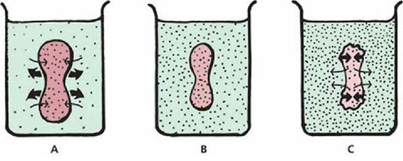
■ FIGURE 2-5 Effect of the tone of a solution on erythrocytes (red blood cells). A. The solution is hypotonic and the erythrocyte expands. B. The solution is isotonic and no change occurs in erythrocyte size. C. The solution is hypertonic and the erythrocyte decreases in size. The thick arrows indicate the direction of cell volume change. The thin arrows indicate the direction of water diffusion.
Table 2-2 presents the results of a laboratory exercise in which erythrocytes from a dog were placed in different concentrations of NaCl solutions. The 0.167 mol/L of NaCl solution (0.977%) was considered isotonic for the erythrocytes of this dog (no change in volume). Both the 0.15 mol/L (0.877%) and 0.10 mol/L (0.585%) solutions were hypotonic (increased volume), while the 0.3 mol/L (1.76%) solution was decidedly hypertonic (decreased volume).
| TABLE 2-2 CHANGES IN VOLUME OF CANINE ERYTHROCYTES ATTRIBUTABLE TO THE TONE OF SUSPENDING NaCL SOLUTION" | ||
| SUSPENDING SOLUTION | VOLUME CHANGE | |
| MOLARITY | PERCENT | PERCENT |
| 0.3 | 1.76 | -16.7 |
| 0.167 | 0.977 | ,0.0 |
| 0.15 | 0.877 | +2.0 |
| 0.10 | 0.585 | +16.7 |
| aValues obtained from student laboratory exercises. | ||
Erythrocytes vary in their ability to withstand hemolysis (rupture of erythrocytes with release of hemoglobin). Older erythrocytes are more fragile and would be the first to hemolyze in solutions with reduced tone. Fragility of erythrocytes can also be increased by certain disease conditions or exposure to toxins and drugs. The degree of fragility can be determined by an osmotic fragility test. Blood from an animal is placed in NaCl solutions with decreasing concentration. The percent hemolysis is determined for each solution in comparison with a solution in which hemolysis would be expected to be 100%. The results of an osmotic fragility test for a normal dog (canine) are presented in Table 2-3 and are compared with those of a normal goat (caprine). It is apparent that goat erythrocytes are less resistant to hemolysis than dog erythrocytes when placed in solutions with increasing hypotonicity. Whereas dog erythrocytes are described as biconcave disks, goat erythrocytes are more spherical; therefore, expansion potential is minimal and hemolysis occurs earlier.
TABLE 2-3 OSMOTIC FRAGILITY OF ERYTHROCYTES FROM NORMAL DOGS
(CANINE) AND NORMAL GOATS (CAPRINE)α
| SUSPENDING SOLUTION | NORMAL.DOGS | NORMAL GOATS |
| PERCENT NaCl | PERCENT HEMOLYSIS | PERCENT HEMOLYSIS |
| 0.85 | 0.0 | 0.0 |
| 0.75 | 0.6 | 2.1 |
| 0.65 | 0.7 | 88.0 |
| 0.60 | 1.7 | 93.6 |
| 0.55 | 14.0 | 97.7 |
| 0.50 | 67.4 | 97.7 |
| 0.45 | 94.4 | 97.7 |
| 0.40 | 95.7 | 100.0 |
| 0.35 | 100.0 | 100.0 |
| 0.30 | 100.0 | 100.0 |
| a Values obtained from student laboratory exercises. | ||
Solutions that cause erythrocytes to enlarge can be sufficiently hypotonic to cause hemolysis of the erythrocytes. Hemoglobin (red in color) in the erythrocyte imparts its color to the solution. Plasma from an animal in which hemolysis has occurred has some degree of redness, depending on the extent of the hemolysis (plasma is usually light yellow to colorless). When this occurs it is known as hemoglobinemia. Sometimes hemolysis occurs to such an extent that hemoglobin enters the kidney tubules and appears in the urine. In this condition, called hemoglobinuria, a red color is imparted to the urine.
Interconversion of Units of Measurement
Solution composition and strength is variably expressed in moles, osmoles, and equivalents, and each has a reference to the weight in grams from which they can be derived. These units are related, and interconversions can be made and must proceed according to the pathways shown in Figure 26.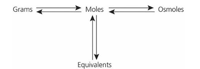
■ FIGURE 2-6 Pathways for the interconversion of grams, moles, osmoles, and equivalents. (From Reece WO. Physicochemical properties of solutions. In: Reece WO, ed. Dukes’ Physiology of Domestic Animals. 13th edn. Ames, IA: Wiley-Blackwell, 2015.)
The problems listed below are frequently encountered when preparing solutions for infusion or when interpreting contents shown on labels for solutions commercially prepared. These problems will enhance your understanding and skill related to physicochemical properties of solutions.
Question 1: How many grams would be needed to prepare 1 L of a 5% glucose solution?
Answer
Step 1: Percent solution = the concentration of solute in grams per 100 mL of aqueous solution. Accordingly, a 5% glucose solution would contain 5 g per 100 mL.
Step 2: Because one liter (1,000 mL) is needed, the amount of glucose would be (5 g ? 1,000) ÷ 100 = 50 g.
Question 2: What is the molarity (M) of.a NaCl solution that contains 8.775 g per L?
Answer:
Step 1: Molarity = g per L/molecular weight (MW).
Step 2: Molecular weight of NaCl = 58.5; therefore, molarity = 8.775/ 58-5 = 0.15 mol/L.
Question 3: What is the osmolarity of a 0.1 mol/L CaCl2 solution?
Answer:
Step 1: Osmolarity is a measure of osmotic pressure and is determined by numbers of particles.
Step 2: One molecule of CaCl2 when placed in solution would ionize and provide three particles (one Ca2+ and two Cl-).
Step 3: The osmolarity (for molecules that ionize in solution) = number ions from molecules ? molarity = 3 ? 0.1 = 0.3 osmol = 300 mosmol (milliosmole).
Question 4: How many grams would be required to make 1 L of a 300 mosmol NaCl solution?
Answer:
Step 1: 300 mosmol NaCl = 150 mmol/L NaCl = 0.15 mol/L NaCl.
Step 2: g/L = molarity ? MW = 0.15 ? 58.5 = 8.775 g.
Question 5: How many equivalents (mEq/L) of Na+ and Cl- are contained in a 0.15 mol/L solution of NaCl?
Answer:
Step 1: NaCl is a monovalent molecule.
Step 2: Eq for each ion = 1 (valence) ? molarity = 0.15 Eq Na+ and 0.15 Eq Cl- = 150 mEq Na+ + 150 mEq Cl-
Question 6: How many equivalents (mEq/L) of Ca2+ and Cl- are contained in a 0.1 mol/L solution of CaCl2?
Answer:
Step 1: CaCl2 is a bivalent molecule.
Step 2: Eq for each ion = 2 (valence) ? molarity = 2 ? 0.1 = 0.2 Eq Ca2+ and 0.2 Eq Cl- = 200 mEq Ca2+ and 200 mEq Cl-.
Question 7: What is the osmolarity (mosmol/L) of a CaCl2 solution labeled to contain 200 mEq Ca2+ and 200 mEq Cl-?
Answer:
Step 1: Convert milliequivalents to millimoles (mEq/valence) = 200/2 = 100 mmol/L CaCl2.
Step 2: Convert mmol/L to mosmol = 100 mmol/L ? number of atoms per molecule (particles) = 100 ? 3 = 300 mosmol.
■