Physiology of the Nerve Impulse
Nerves rapidly transmit information from one body site to another via nerve impulses (i.e., action potentials propagated along the axons of neurons within the nerves). The genesis of a resting membrane potential and the development of action potentials and their propagation are described in detail in Chapter 2 and are only briefly reviewed here.
The resting membrane potential of neurons depends mainly on nongated potassium channels in the cell membrane and the electrogenic Na—K—ATPase, or Na—K, pump. The continuous exit of potassium from the cell down its concentration gradient and the continuous activity of the pump maintain a difference in charge across the membrane so that the interior is negative relative to the exterior (i.e., resting membrane potential).
The axonal cell membranes of neurons contain voltage-gated sodium channels. These channels are closed at normal resting membrane potentials but rapidly open when the membrane potential is brought to threshold voltage. This results in the inward movement of sodium (down its concentration gradient) and a large, rapid membrane depolarization (Fig. 10-1). At the peak of the action potential (maximal depolarization), the sodium channels close and some additional voltage-gated potassium channels are open. The
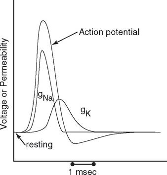
Figure 10-1. Change in sodium and potassium permeability during neuron action potential. The lines labeled gNa and gK show the respective changes in membrane permeabilities for sodium and potassium, and the changes are drawn to illustrate their time course relative to the action potential.
closing of the sodium channels and the exit of additional potassium repolarizes the membrane. The exit of additional potassium produces a small afterhyperpolarization until resting conditions can be reestablished (Fig.
10-1).When an action potential occurs on the axon of a neuron, the membrane potential of adjacent areas is altered by local movement of charge. Normally, this causes the sodium channels in the adjacent area to reach their threshold voltage, and another action potential is elicited. By this means action potentials can be propagated along axons (Fig. 2-16). Propagation normally occurs in only one direction, in part because the sodium channels where the action potential just occurred are refractory to another stimulus. This refractory period is a characteristic of normal sodium channels, and the channels rapidly pass through this period so that they are no longer refractory when another normal impulse arrives.
Conduction Velocity and Myelination
As described in Chapter 9, the axons of neurons may be either unmyelinated or myelinated.
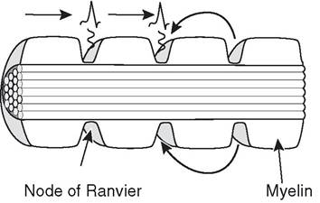
Figure 10-2. Propagation of the action potential in a myelinated axon. Action potentials occur at each node of Ranvier.
Unmyelinated axons have voltage-gated sodium channels throughout their cell membrane, and thus the propagation of action potentials along unmyelinated axons is as described earlier. The myelin sheath that surrounds myelinated axons contains the lipid sphingomyelin, which is a good insulator against ionic flows. Each Schwann cell covers about a 1-mm distance along the axon, and the junction between two schwann cells is called the node of Ranvier. No myelin is present at these nodes, and voltage-gated sodium channels are especially prevalent in the axonal cell membrane at the node. Therefore, the circuits of current must flow from node to node, and action potentials occur only at the nodes. since this is analogous to jumping from one node to the next, it is given the name saltatory conduction (from Latin saltare, to jump) (Fig. 10-2). This type of conduction contributes to the increased rate of impulse conduction in myelinated axons.
Conduction velocities of axons also depend on their diameter. Large-diameter axons propagate action potentials at higher velocities than do small-diameter axons, because large axons have less internal resistance to the flow of current. Large myelinated fibers, 20 μm in diameter, approach conduction velocities of 250 mph (130 m/sec). At this rate an impulse could travel 6 feet in about 15 m/sec. The smallest unmyelinated fibers of the body, about 0.5 μm in diameter, conduct at only about 20 inches per second (0.5 m/sec).