PITUITARY GLAND
1. What is the hypophysioportal circulation? What is its function?
2. What are the abbreviated names of the anterior pituitary hormones?
3. Briefly list the functions of each of the anterior pituitary hormones.
Is STH needed throughout life or only during the growth phase?4. What is meant by posterior pituitary hormones being known as neuro secretions?
5. Briefly list the functions for the posterior pituitary hormones.
The pituitary gland (hypophysis cerebri) has two distinct parts, the anterior lobe (anterior pituitary; adenohypophysis) and the posterior lobe (posterior pituitary; neurohypophysis). It is located in a bony recess (sella turcica) at the base of the brain. The divisions, blood supply, and neural connections to the hypothalamus are shown in Figure 6-1. Its location just below the hypothalamus provides for direct delivery of releasing and inhibiting hormones from the hypothalamus to the anterior lobe and for direct entry of secretory neurons from the hypothalamus to the posterior lobe. Assisting the delivery of hormones to the anterior lobe is a unique arrangement of blood vessels, the hypophysioportal circulation (see Figure 6-1). Similar to other blood portal systems, the venous blood drained from the hypothalamus is redistributed by another capillary system within the anterior lobe. Shortages of hormones in arterial blood are detected by specific cells within the hypothalamus, which are stimulated to secrete releasing hormones. The hormones produced are distributed by the second capillary bed to their appropriate cells in the anterior lobe.
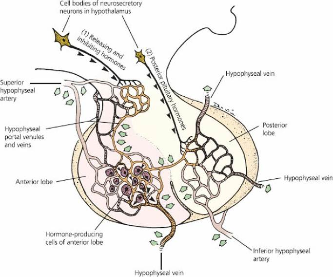
■ FIGURE 6-1 Schematic representation of the pituitary gland and its hypophysioportal circulation. Hypothalamic releasing and inhibiting hormones (1) reach anterior pituitary cells by way of this circulation (left).
Posterior pituitary hormones (2) enter capillaries of the posterior pituitary (right). Open arrows indicate direction of blood flow; arrowheads indicate direction of hormone transport toward axon terminals. (From Cormack DH. Essential Histology. 2nd edn. Baltimore, MD: Lippincott Williams & Wilkins, 2001.)Anterior Pituitary
The anterior pituitary lies forward from the posterior pituitary and has five different cell types that secrete the seven hormones of the anterior pituitary: (1) somatotrope cells, which secrete growth hormone; (2) corticotrope cells, which secrete adrenocorticotropic hormone and beta-lipotropin hormone; (3) mammotrope cells, which secrete prolactin; (4) thyrotrope cells, which secrete thyroid stimulating hormone; and (5) gonadotrope cells, which secrete follicle-stimulating hormone and luteinizing hormone.
Because of the relatively large number of important hormones associated with the pituitary gland, it is sometimes called the master gland. In the past, pharmaceutical companies obtained animal pituitary glands from slaughter houses and extracted several hormones for commercial and experimental uses. Recovery of the pituitary gland at slaughter is laborious because of its protected location, and yields are low (340 g/100 cattle; 30 g/100 pigs) because of its small size.
Anterior Pituitary Hormones
The hormones of the anterior pituitary belong to the peptide class, ranging from polypeptides to large proteins. Differences in structure are noted among species and replacement therapy from one species to another is not uniformly successful. Sometimes an active core of a hormone is identified that permits its subsequent use after the noncore portion is removed.
Growth Hormone
Growth hormone is also known as somatotropic hormone (STH) because of its stimulating effect on the somatic cells (body cells). It has invariably been referred to as growth hormone because of its stimulation of increase in body size. It causes growth of all tissues of the body that are capable of growth and it promotes both increased cell size and increased mitosis with development of increased cell numbers.
The epiphyseal plates of long bones are particularly sensitive to growth hormone; it stimulates mitotic activity, which results in lengthening. Growth hormone stimulates the liver to form several small proteins called somatomedins, which then act on cartilage and bone to promote their growth. Bone and cartilage are therefore not stimulated directly by growth hormone, but indirectly by this intermediate compound.In addition to its general effect of causing growth, STH has several specific metabolic effects. Because of these, it is apparent that STH is needed throughout life and not only during the growth phase. These metabolic effects include: (1) increased rate of protein synthesis in all body cells, (2) increased mobilization of fatty acids from fat and increased use of fatty acids for energy, and (3) decreased rate of glucose uptake throughout the body. The preferential use of fats for energy conserves glucose and promotes glycogen storage. Because of glycogen storage, the heart can endure emergency contraction more effectively, whereby glycogen stored in the heart is converted to glucose. Probably most metabolic functions of growth hormone are caused not by its direct effects on the tissues but by indirect effects through the somatomedins.
An effect of growth hormone in increasing milk yields in the lactating cow has received considerable research interest. Growth hormone does not produce its effects by stimulation of the mammary gland, but, rather, it seems that the increased milk yield caused by continuous injections of exogenous STH is caused by the partitioning of available nutrients from body tissues toward milk synthesis.
Adrenocorticotropic Hormone
Adrenocorticotropic hormone (ACTH) causes increased activity of the adrenal cortex. It was formerly thought that ACTH only stimulated the secretion of glucocorticoids by the adrenal cortex, but it is now recognized that mineralocorticoid (aldosterone) secretion is also enhanced. In addition, it has become apparent that ACTH has metabolic effects somewhat similar to those of STH, in which protein synthesis and fatty acid uptake are enhanced and glucose uptake is decreased.
Thyroid-Stimulating Hormone
Thyroid-stimulating hormone (TSH) stimulates the synthesis of colloid by thyroid gland cells and stimulates the release of thyroid hormone. Associated with these functions are the accumulation of iodine, organic binding of iodine, and formation of thyroxine within the thyroid gland. No extrathyroid activity is apparent for TSH, as for STH and ACTH.
Gonadotropic Hormones and Prolactin
The gonadotropic hormones, follicle-stimulating hormone (FSH) and luteinizing hormone (LH), have specific roles in male and female reproduction, and detailed accounts are provided in Chapter 14 (Male Reproduction) and Chapter 15 (Female Reproduction). Specifically, FSH stimulates oogenesis and spermatogenesis in the female and male, respectively. In the female, LH assists ovulation and development of a functioning corpus luteum and in the male it stimulates the secretion of testosterone. Prolactin helps to initiate and maintain lactation after pregnancy. Also, in the ewe, it is associated with maintenance of the corpus luteum.
Beta-Lipoprotein Hormone
Beta-lipoprotein hormone (β-LPH) is secreted by the same cells (corticotrope) that secrete ACTH (see previous text). The physiologic role of β-LPH is still unknown. Products providing pain relief (e.g., endogenous opiates, which are the endorphins and enkephalins) might be derived from β- LPH. Inasmuch as they are associated with ACTH (same secreting cells), the response to stress might include β-LPH secretion and pain relief as a neural response.
Posterior Pituitary and Its Hormones
The posterior lobe is an outgrowth of the hypothalamus (see Figure 6-1) and contains the terminal axons from two pairs of nuclei (supraoptic nucleus and paraventricular nucleus) located in the hypothalamus. The supraoptic and paraventricular nuclei synthesize antidiuretic hormone and oxytocin (neurosecretions), respectively, which are transported to the axon terminals in the posterior pituitary, where they are stored in secretory granules until released.
An action potential, generated by the need for each of the stored hormones, causes the release of the hormone and subsequent absorption into the blood, where it is distributed to the receptor cells. The hormones of the posterior pituitary are of the peptide class, specifically nonapeptides (they contain nine amino acids).Antidiuretic Hormone
When an animal is given an overload of water, a period of diuresis (increased output of dilute urine) occurs. Diuresis can be prevented by the administration of antidiuretic hormone (ADH), also known as vasopressin. If dehydration occurs (osmoconcentration), osmoreceptors respond to the increased concentration by stimulating greater output of ADH by the axon terminals in the posterior pituitary. The target cells of the secreted ADH are the collecting tubules and the collecting ducts of the kidney. The presence of ADH renders the cells of the collecting tubules and collecting ducts more permeable to water, and more water is absorbed from the tubular fluid so that the plasma osmolality decreases (Na+ concentration returns to normal) and the urine volume decreases (becomes more concentrated). ADH is therefore important for water conservation by animals. Other stimulators of ADH secretion include reduced blood volume, trauma, pain, and anxiety.
Oxytocin
The functional activity of oxytocin is related to the reproductive processes, which include lactation (see Chapter 16 ). Oxytocin is released from the posterior pituitary as a result of neuroendocrine reflexes. The act of suckling or similar teat stimulation causes the release of oxytocin and subsequent milk letdown. Similarly, an estrogen-dominated myometrium, such as is found at ovulation and at parturition, is more responsive to oxytocin, and greater contraction of the uterus results. Oxytocin release at these times is associated with appropriate stimuli and subsequent myometrial contraction, which assists in the transport of sperm to the oviduct at copulation and in the expulsion of the fetus at parturition.