THYROID GLAND
1. What is the substance that fills the thyroid follicles?
2. Sketch the thyroxine molecule and note the presence of iodine. How does T3 differ from T4?
3.
What is thyroglobulin? How are T3 and T4 stored in the thyroid gland after their formation? Describe the release and absorption of T3 and T4 from the thyroid follicles.4. What fraction of thyroid hormone release from the thyroid gland is T4?
5. Describe the plasma transport, release, and cell use characteristics of T3 and T4.
6. What is the most well-known function of the thyroid hormones?
7. Note how low levels of thyroid hormones cause the secretion of the thyroid hormones.
8. What is calcitonin? Where is it secreted? Is it secreted in response to hypercalcemia or hypocalcemia? What, then, is its function?
In most mammals the thyroid gland is located on the trachea, just caudal to the larynx. In cattle it consists of two laterally placed, somewhat flattened, lobes joined by an isthmus (Figure 6-2). The lateral lobes have a less substantial isthmus in the horse and no isthmus in the dog and cat. Pigs have a compact thyroid form with a large median lobe (instead of an isthmus) in addition to the lateral lobes. The thyroid gland is composed of numerous follicles (Figure 6-3) lined by simple cuboidal epithelial cells and filled with a fluid known as colloid. The surface area of the lining epithelium is increased by villi that project into the follicle.
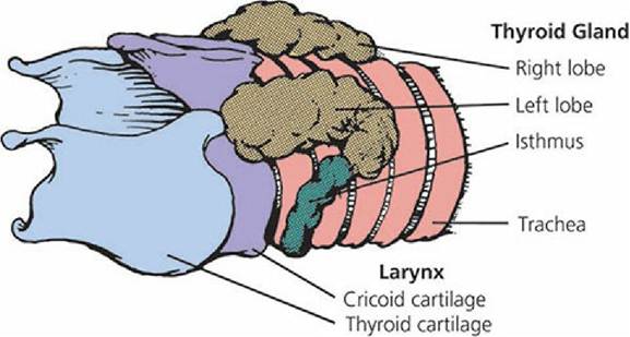
■ FIGURE 6-2 Thyroid gland (bovine).
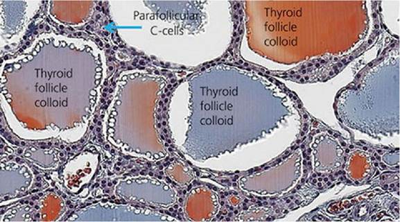
■ FIGURE 6-3 Photomicrograph of thyroid follicles showing iodinated thyroglobulin stored within the colloid. Parafollicular cells (C cells that secrete calcitonin) are positioned beside follicles and usually occur as single cells (arrow).
(From Goff JP. The Endocrine System. In Reece WO. ed. Dukes’ Physiology of Domestic Animals. 13th edn. Ames, IA: Wiley-Blackwell, 2015.)Thyroid Hormones
The thyroid hormones belong to the amine classification of hormones - they are derived from the amino acid tyrosine. A further characteristic of the thyroid hormones is that they contain iodine. Iodine is bound organically in the thyroid gland in four forms. The thyroid hormones, 3,5,3',5'- tetraiodothyronine (thyroxine [T4]) and 3,5,3'-triiodothyronine (T3) (Figure 6-4), are combinations of two molecules of 3,5-diiodotyrosine, as in T4, or one molecule of 3-monoiodotyrosine with one molecule of 3,5-diiodotyrosine, as in T3. In both cases the combination results in the loss of one water molecule and an amino acid residue, glycine. Iodine trapping and iodination are unique features of the thyroid gland that are assisted by TSH.
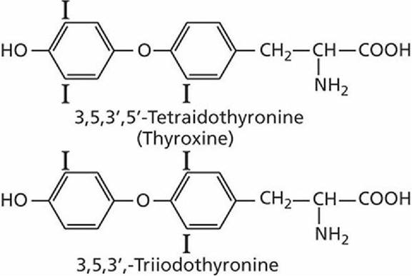
■ FIGURE 6-4 Structural formulas of the thyroid.hormones, thyroxine (T4) and 3,5,3'- triiodotyrosine (T3).
Biochemistry of T3 and T4 Formation
Thyroglobulin is a large glycoprotein molecule secreted into the follicle by the lining cells.
Thyroglobulin has a molecular weight of about 680,000. It contains many tyrosine molecules; when iodinated, they consist of both 3-monoiodotyrosine and 3,5-diiodotyrosine.
Tyrosine coupling occurs while the tyrosine residues are still attached to the thyroglobulin molecule. The lining cells of the follicles provide the enzymes required for coupling to form T3 and T4. The coupled tyrosines, still attached to the thyroglobulin molecule, are stored after synthesis within the follicle.
Release and Transport of T3 and T4
The thyroglobulin molecule, with enclosed T3 and T4, is not released into the blood from the thyroid follicles. Extensions from the follicle cells enclose parts of colloid so that colloid becomes a vesicle within the cell (endocytosis).
Lysosomes release proteolytic enzymes that separate T3 and T4 from thyroglobulin and permit their absorption from the base of the cells. The 3-monoiodotyrosine and 3,5-diiodotyrosine freed similarly by digestion are not absorbed, but are deiodinated, and both the iodine and the tyrosine are recycled into new thyroglobulin. About 90% of thyroid hormone released is T4.T3 and T4 are combined immediately with plasma proteins for transport in the blood. The major plasma protein is termed thyroxine-binding globulin (TBG). This protein has a high affinity for the thyroid hormones, but a greater affinity for T4 than for T3. All of the T4 and T3 are bound, but because of the greater affinity of TBG for T4 than for T3, more T3 is released to tissue cells than T4. Once within the tissue cells, T3 is more potent than T4, but its duration of action is shorter. Thus, short-term and long-term demands by cells can be met effectively by the different release and potency characteristics associated with T4 and T3.
Functions
The most well-known function of thyroid hormones is their ability to increase internal heat, thereby increasing the rate of oxygen consumption. Thyroid hormones stimulate the metabolic activities of most tissues of the body except for the brain, lungs, retina, testes, and spleen. The ability to increase metabolic activity and oxygen consumption is partly a result of the stimulation or activation of some key enzymes, including alpha-glycerophosphate dehydrogenase, hexokinase, diphosphoglycerate mutase, and cytochromes b and c. The lipolytic effect of epinephrine is also markedly potentiated by thyroid hormones. The specific role of thyroid hormones in increasing internal heat production has not been clearly defined, but it has been suggested that the increased heat is secondary to increased protein synthesis stimulated by thyroid hormones.
Regulation of Secretion
To provide consistent quantities of thyroid hormones, feedback control mechanisms are provided through the hypothalamus and anterior pituitary.
Decreased levels of thyroid hormones result in the secretion of thyrotropin-releasing hormone (TRH) into the hypophysioportal circulation. The thyrotrope cells of the anterior pituitary are thereby stimulated by TRH to secrete TSH. TSH secretion,is followed by increased thyroid gland activity, including the release of T3 and T4,from the thyroglobulin molecule, their absorption into the blood, and transport to cells. A constant level consistent with normal metabolism is ensured by this feedback mechanism. The rate of TSH secretion increases above normal by the exposure of animals to cold environments. The response is mediated by the cooling of the anterior hypothalamus, resulting in an increase in metabolic rate and an attendant increase in heat production. Excitement and anxiety result in a decreased output of thyroid hormones because the stimulation of the sympathetic nervous system by these states causes an increase in epinephrine and norepinephrine output, resulting in an increase in metabolic rate and heat production.Thyroid Deficiency and Antithyroid Compounds
The typical form of thyroid hormone deficiency results from an iodine deficiency and consequent inability of the thyroid gland to produce T3 and T4. The lack of circulating hormones causes the usual feedback mechanisms so that TSH is produced, and the resulting stimulation of the thyroid gland causes thyroglobulin accumulation without effective output of T3 and T4. The thyroid gland enlarges because of colloid accumulation, a condition known as goiter. Thyroid gland enlargement can be caused by hypothyroidism (e.g., iodine deficiency) or hyperthyroidism (e.g., increased thyroxine demands, tumor). Goiter caused by iodine deficiency is rarely seen in the domestic animals and other causes of thyroid dysfunction are relatively uncommon in sheep, cattle, and swine. The clinical signs of hypothyroidism and hyperthyroidism, however, are common in dogs and cats. Lack of activity (lethargy), hair loss, dry and dull hair, cold sensitivity, and anemia are common clinical signs of hypothyroidism.
Fatigue, weight loss, hunger, nervousness, and sensitivity to heat are associated with hyperthyroidism.Natural substances have been identified that cause goiter by inhibiting thyroid function, called goitrogens. Because thyroid function is inhibited, thyroxine is not produced in sufficient amounts and TSH continues to be secreted, resulting in thyroglobulin accumulation. One such goitrogen, goitrin, is produced in the intestinal tract after the ingestion of a progoitrin contained in cruciferous plants (e.g., cabbage, rutabaga, turnip). Thiocyanate, another goitrogen, is also contained in some plants, and plant goitrogens are important causes of animal goiter in some parts of the world. Goitrin and related compounds cause goiter by interfering with the organic binding of iodine, but thiocyanate interferes with iodine trapping by the thyroid gland. In the latter case, the effects can be overcome by feeding excess iodine. Antithyroid compounds are used for the treatment of hyperthyroidism; these include methimazole, propylthiouracil, and carbimazole. The use of antithyroid compounds to promote weight gain has not produced satisfactory results.
Calcitonin Calcitonin is a hormone of the thyroid gland secreted by parafollicular or C cells (see Figure 16-3) that are also present in the walls of thyroid gland follicles. Calcitonin is a polypeptide of 32 amino acids (MW 3,000).
The stimulation for secretion of calcitonin is hypercalcemia and, to a lesser extent, hypermagnesemia. Calcitonin inhibits osteoclastic bone resorption (see Chapter 7) and thereby attempts to lower the plasma Ca2+ concentration. Calcitonin also inhibits phosphate resorption and enhances calcium loss in the kidney. Calcitonin is antagonistic to the action of another hormone associated with calcium homeostasis, parathyroid hormone (see later section). The latter hormone protects against low plasma concentrations of Ca2+.
■