Prenatalgrowth
Prenatal growth starts when zygote is formed and continue till the birth of animal. This includes a series of orderly differential processes that transform a single celled zygote into multicellular organism (Figure 23.2).
23.7.1 Three Stages of Prenatal Growth
23.7.1.1 Cleavage
Division and re-division of cells without an increase in the volume of cytoplasm (Figure 23.3).
The cleaving cells are known as blastomeres. Cleavage is characterized by a progressive increase in cell number but a decrease in cell size. Nuclear multiplication involves the synthesis of significant amounts of DNA, which is one of the main constituents of chromosomes and chromatin. The early cleavages are synchronous and occur at specific times after fertilization. During later cleavages, cell division is asynchronous since different parts of the embryo cleave at different rates. When the dividing cells reach a certain number characteristic of the species, the cells start pushing each other due to the accumulation of fluid. The embryo, then called a blastocyst, which is a spherical or flattened hollow ball. The cavity developed inside is called
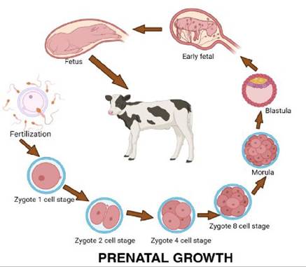
FIGURE 23.2 Prenatal growth
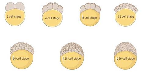
FIGURE 23.3 Cleavage of cells
blastocoelic cavity. The blastomeres vary in size, yolk content, and cytoplasmic organization, but until this stage, no organ formation starts.
23.7.1.2 Differentiation
Progressive specialization of cells both structurally and functionally. During the 1st week of gestation, the blastocyst differentiates into two types of cells: a single layer of outermost cells known as the trophoectoderm/trophoblast and a small aggregation of cells beneath the trophoecto- derm known as inner cell mass (ICM)/embryonic disc.
The trophoectoderm leads to the establishment of the placenta, whereas inner cell mass develops into the embryo proper.The blastocyst then further differentiates into gastrula, which is a three-layered structure
• Outermost layer: Ectoderm
• Middle layer: Mesoderm
• Innermost layer: Endoderm
These three layers are also known as germinal layers/germ layers. These three germinal layers give rise to the organs of the embryo.
23.7.1.3 Organogenesis (Organ Formation)
Organogenesis starts when the three germinal layers are formed at the gastrula stage. It occurs by:
• Invagination (in folding)
• Envagination (out folding)
• Budding
• Hollowing out
Cells from each of the three germ layers aggregate into primordial cell masses from which special organs will be formed. The outermost layer or ectoderm forms a mid-dorsal ridge along the antero-posterior axis of the blastocyst quite early in development. This elongated ridge of neural ectoderm subsequently forms the brain, spinal cord, and other derivatives of the nervous system. Ectodermal cells lateral to the neural ectoderm develop into the skin and its derivatives such as mammary glands, sweat glands, sebaceous glands, hair, nails, and hoofs.
Innermost layer (endoderm) forms:
• Inner lining of the gut and its glands,
• Liver and pancreas
• Lining of trachea, bronchi and lungs
• Thyroid, parathyroid and thymus gland
• Urinary bladder
Middle layer (mesoderm) gives rise to:
• Bones and muscles
• Reproductive and excretory system
• Blood and blood vessels
• Inner layer of skin/dermis
Organogenesis completed in 35 days in cows, 40 days in horses, and 60 days in humans.
Most organs are not functional when they first appear in the embryo. The embryonic structures start functioning only after further differentiation. The differentiation and development of embryonic and fetal structures are called developmental horizons and are useful criteria for determining the age of embryos and fetuses.
For example, in cattle, the earliest formation of most organs and body parts occurs during the 2nd - 6th week of gestation. The digestive system, lungs, liver, and pancreas develop from the primitive gut during this period.23.7.1.4 Fetal Growth
Once the organogenesis is completed, the embryo is now called a fetus. After organogenesis, there is a period of rapid increase in the organ size of fetus (Figure 23.4).
This increase in dimensions of the fetus is a result of cell proliferation (Hyperplasia) and an increase in cell size (Hypertrophy). This is called fetal growth. The phases of cellular differentiation in which the fetal tissues develop their definite characteristics are known as histogenesis (e.g., cells which become muscle, develop contractile fibrils). The rate of growth in early stages of fetal development depends upon:
Genetic make up which is variable among species. In general, the growth rate is faster in smaller species than in larger ones.
Supply of adequate nutrition. Nutrition has a major influence on growth. It should be adequate in the early stage of fetal development so that the fetus becomes larger and gains considerable weight per day.
Maternal blood supply. The mother’s blood supplies the fetus with all its nutrients. The quantity of blood reaching the fetus determines its rate of growth.
Variation in Prenatal Growth. There are many variations in the prenatal growth of different species that might be due to their different genetic make up, developmental process and gestation period
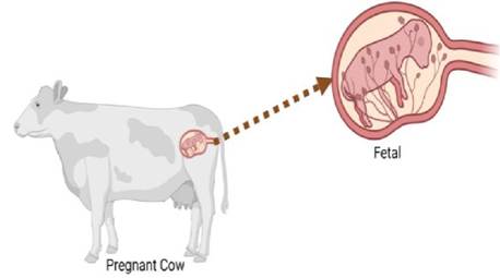
FIGURE 23.4 Fetal growth
In sheep, a lamb is born with about the same weight as a human infant but the gestation period is only half as long (150 days vs. 280 days). The reason being is that the fetal lamb grows more rapidly than the human fetus during the first stage of development, and this difference is maintained throughout its development in utero.
In rats, during first 3 weeks of growth, the increase in the number of cells goes from single cell to 2-3 million cells. By the time when it leaves the uterus, it weighs about 5 g, while in humans, in the same time period from conception, a human fetus attains only about one-hundredth of this weight and number of cells.
Small rodents (mouse, rat, rabbit, etc.) grow extremely rapidly, while cat, dog, pig, goat, sheep, cattle, horses, etc., grow less rapidly in the early stages. But these species grow for a longer time in the uterus, so they grow larger in size than rat.
23.7.2 Growth of Fetus Occurs Differentially
The birth weight of the fetus = approx. 60% of the total weight of the conceptus. The fetal membranes (amnion, chorioallantois, and chorionic villi) and placental fluids (amniotic and allantoic) make up the remaining 40%. Different parts of the conceptus grow at different rates during gestation. For example, during the early stages of gestation, the fetal membranes grow rapidly, and their absolute weight reaches a maximum during mid-pregnancy. The increase in the volume of placental fluids corresponds more or less to that of the fetus. Toward the end of gestation, the placental fluids tend to decrease in volume, although the fetus is still growing.
In the early stage of embryonic development, the cephalic region grows rapidly, so that the head of the fetus is disproportionately large. During the later stages of fetal life, when growth in the cephalic region is less rapid, the proportions of the neonate are established. The growth gradients of the fetus follow a definite order (e.g., the head, limbs, and the forequarter are relatively more developed than other parts of the body at the time of birth). Since the various organs of the fetus grow at vastly different rates, the conformation of the fetus is continuously changing.
23.7.3 Factors Affecting Prenatal Growth
Heredity/genetics: Genes influence the growth of an individual animal.
The maternal contribution is more important than the paternal, which is directly affects litter size and fetal size. Further variations between species, strains, etc., also depend upon genetic set up.Size of dam: The faster prenatal growth is directly related to larger maternal size. Walton and Hammond conducted experiments on reciprocal crosses between two different breeds of horses: - Large Female X Small Male (Shire X Shetland) resulted in foals bigger in size (Shire) (Shetland)
Larger Male X Small Female resulted in foals smaller in size
This difference in size was maintained even up to 4 years of age. The smaller maternal environment restricts the growth of the fetus, making parturition easier. A small placenta restricts its growth.
Age of dam: A young mother that is not fully developed during pregnancy competes with the fetus for available nutrients, thus not let the fetus to grow to its full potential.
Parity of dam: Young ones born to aged females are also smaller, because there is a lot of fat deposition around the uterus, which restricts further growth of the young one. (Less space availability).
Maternal nutrition: The fetus continues to grow up to near normal with poor nutrition during early pregnancy. However, young ones born to poorly fed dams during late pregnancy show stunted growth and these mothers may give birth to premature young ones.
In sheep/cattle: if the animal during early pregnancy but properly during late pregnancy, there is no effect on fetal growth, the birth weight is also normal. But if poorly fed during late pregnancy, lambs have stunted growth.
During last stage of pregnancy the variation in fetal weight reflects the differences in genetic factor, litter size, nutritional status, and health of the dam. When the mother is adequately fed, the birth wt. is towards the upper limit (male fetus grow more than the female). Poor nutrition of mother affects glycogen content of the fetal muscle and liver that may be the cause of neonatal mortalities.
Fetal glycogen uses as a source of energy immediately after birth and normally builds up during later stage of pregnancy. Restriction of diet of mother produces differences in growth of different organs of fetus body. The nervous system, skeletal system and heart are least affected, whereas kidneys, lung and muscles are moderately affected. The skin, thymus, spleen and liver are highly affected organ of fetus.Litter size: In polytocous species, increase in litter size retards the prenatal growth. Similarly, in monotocous species, multiple births usually retard prenatal growth but the combined weight of two or more fetus is more than the single fetus. This is due to variation in function of placenta. The placenta determines the prenatal growth. Smaller growth with increase in litter size is mainly because lesser nutrients reach each fetus. Thus, a small fetus could be due to Inherent low growth potential and Poor supply of blood, nutrients from mother to fetus.
Placenta size: Smaller size of placenta retards the size of fetus as the nutrients pass to the fetus through the placenta. The placenta acts as a blood reservoir. So bigger the placenta more blood (nutrients) reach the fetus. When there is large no. of placenta in one uterine horn, it decreases the flow of blood and oxygen supply to the distally positioned fetus/ placenta which leads to intra uterine transfusion syndrome which markedly affects the prenatal growth.
Ambient temperature: Stunted prenatal growth is observed when pregnant animals exposed to high temperature. Degree of stunted growth of fetus in uterus depends on the length of exposure to the environmental temperature.
Fetal hormones: Fetal hormones regulate functioning of the fetus. When the levels of these hormones are not sufficient they may affect the fetal growth, e.g. Growth hormone, thyroid hormones, adrenal hormones etc. GH regulates metabolic processes in the growing fetus while thyroxin and androgens are required for morphogenesis. In man and rat, the brain fails to develop normally when thyroxin is deficient. Morphogenesis of external genitalia (e.g. scrotum and penis) is androgen dependent. In absence of fetal androgens, morphogenesis of external genitalia fails to occur and external genitalia will not develop properly.
23.8