RECEPTORS OF NEUROTRANSMITTERS
Neurotransmitters of PNS and CNS, either excitatory or inhibitory, exert their effects by binding to specific receptors on the postsynaptic neuron or other target cells.
• Based on the structure, there are two main types of neurotransmitter receptors:
1.
Ionotropic receptors (ligand-gated ion channels): These receptors directly regulate the flow of ions across the cell membrane when bound by neurotransmitters. When a neurotransmitter binds to an ionotropic receptor, it causes a conformational change in the receptor protein, leading to the opening or closing of an ion channel. This rapid change in ion flow directly alters the membrane potential of the postsynaptic neuron, either depolarizing it (excitatory effect) or hyperpolarizing it (inhibitory effect). Examples include nicotinic acetylcholine receptors and certain glutamate receptors.2. Me tabotropic receptors (G protein-coupled receptors): These receptors indirectly influence ion flow and cellular processes through intracellular signaling pathways when activated by neurotransmitters. When a neurotransmitter binds to a metabotropic receptor, it activates a G protein, which then initiates a cascade of intracellular signaling events. These signaling pathways can modulate the ion channels, and typically induce slower and longer-lasting effects compared to ionotropic receptors. Examples include muscarinic acetylcholine receptors, dopamine receptors, and serotonin receptors.
• Based on the neurotransmitter that the receptors bind to, they are broadly classified into: Adrenergic, Cholinergic and Glutamate receptors. The salient features of adrenergic, Cholinergic and Glutamate receptors are tabulated in Table 8.4.
TABLE 8.4
Salient Features of Adrenergic, Cholinergic and Glutamate Receptors
| Feature | Adrenergic Receptors | Cholinergic Receptors | Glutamate Receptors |
| Types/Subtypes | Alpha (α1, α2) and Beta (β1, β2, β3) | Nicotinic (nAChR) and Muscarinic (mAChR) | Ionotropic (AMPA, NMDA, Kainate) and Metabotropic (mGluR) |
| Primary Ligand | Norepinephrine (noradrenaline) and Epinephrine (adrenaline) | Acetylcholine (ACh) | Glutamate |
| Receptor Mechanism | G-protein coupled receptors (GPCRs) | nAChR: Ligand-gated ion channels, mAChR: GPCRs | Ionotropic: Ligand-gated ion channels, Metabotropic: GPCRs |
| Location | Various tissues, including heart, lungs, blood vessels, CNS | Neuromuscular junction, autonomic ganglia, CNS, various tissues | CNS, peripheral nervous system, various tissues |
| Primary Functions | Regulation of cardiovascular functions, smooth muscle tone, metabolic processes | Neuromuscular transmission, autonomic control, various CNS functions | Synaptic transmission, neural plasticity, excitatory neurotransmission |
| Response Time | Relatively slow (milliseconds to seconds) | nAChR: Fast (milliseconds), mAChR: Slow (milliseconds to seconds) | Ionotropic: Fast (milliseconds), Metabotropic: Slow (seconds) |
| Common Agonists | Epinephrine, Norepinephrine | Nicotine (nAChR), Muscarine (mAChR) | Glutamate, NMDA (for NMDA receptors), AMPA (for AMPA receptors) |
| Common Antagonists | Alpha-blockers (e.g., phentolamine), Beta-blockers (e.g., propranolol) | nAChR antagonists (e.g., curare), mAChR antagonists (e.g., atropine) | NMDA receptor antagonists (e.g., ketamine), AMPA receptor antagonists |
TABLE 8.5
Differences Between Nicotinic and Muscarinic Receptors
| Feature | Nicotinic Receptors |
| Type | Ionotropic (ligand-gated ion channels) |
| Primar location | Neuromuscular junctions, autonomic ganglia, CNS |
| Subtypes | N1 (NM) - muscle type, N2 (NN) - neuronal type |
| Agonists | Nicotine |
| Antagonists | Curare, hexamethonium |
| Mechanism of action | Directly opens ion channels, allowing Na+ and K+ to pass |
| Response time | Fast (milliseconds) |
| Physiological effects | Skeletal muscle contraction, autonomic ganglionic transmission |
| Synaptic transmission | Excitatory |
| Clinical relevance | Used in anesthesia (neuromuscular blockers) |
Muscarinic Receptors
Metabotropic (G-protein coupled receptors)
CNS, parasympathetic nervous system (heart, smooth muscles, glands)
M1, M2, M3, M4, M5
Muscarine
Atropine, scopolamine
Activates G-proteins, leading to various intracellular effects
Slow (seconds to minutes)
Modulation of heart rate, smooth muscle contraction, glandular secretion
Can be excitatory or inhibitory
Used in treating various conditions like bradycardia, asthma, and gastrointestinal disorders
8.16.1 Cholinergic Receptors
Cholinergic receptors are receptors that bind the neurotransmitter acetylcholine and mediate its effects on target cells.
There are two main types of cholinergic receptors. The differences between nicotinic and muscarinic cholinergic receptors are given in Table 8.5.1. Nicotinic acetylcholine receptors (nAChRs): Nicotinic receptors are pentameric ion channels composed of five subunits arranged around a central pore. They are named after nicotine, a compound found in tobacco which mimics the effects of ACh on these receptors, leading to their activation. Binding of ACh or nicotine to the receptor causes conformational changes that open the channel, allowing the passage of cations Na+ and K+. This results in depolarization of the postsynaptic membrane, leading to excitatory effects. Nicotinic receptors are widely distributed in both CNS and PNS. In the CNS, they are primarily found at synapses between neurons and are involved in fast synaptic transmission. In the PNS, nicotinic receptors are found at neuromuscular junctions, where they mediate the effects of ACh on skeletal muscle contraction, as well as in autonomic ganglia, where they mediate synaptic transmission between preganglionic and postganglionic neurons of the autonomic nervous system. Activation of nicotinic receptors can lead to various physiological responses, including muscle contraction, release of neurotransmitters in the brain, and modulation of synaptic transmission. Their effects on different tissues are given in Table 8.6.
2. Muscarinic acetylcholine receptors (mAChRs): Muscarinic receptors are metabotropic receptors that bind with acetylcholine and are named after
TABLE 8.6
Action of Nicotinic Cholinergic Receptors in Different
Tissues
| TissueZLocation | Action of Nicotinic ACh Receptors |
| Neuromuscular Junction | Stimulation of skeletal muscle contraction |
| Autonomic Ganglia | Stimulation of sympathetic and |
| parasympathetic ganglia | |
| Adrenal Medulla | Stimulation of catecholamine release |
| (epinephrine and norepinephrine) | |
| Brain | Modulation of neurotransmitter release and |
| neuronal excitability | |
| Peripheral Nerves | Modulation of neurotransmitter release and |
| neuronal excitability |
muscarine, a compound found in certain mushrooms that activates these receptors. Unlike nicotinic receptors, which are ionotropic and directly regulate ion flow across the cell membrane, muscarinic receptors indirectly modulate cellular processes through intracellular signaling pathways when activated by ACh.
Muscarinic receptors are G protein-coupled receptors (GPCRs) having five subtypes, designated M1 through M5, which are encoded by different genes and exhibit distinct tissue distributions and signaling properties. In the CNS, muscarinic receptors play roles in regulating synaptic transmission, modulating neuronal excitability, and mediating various cognitive functions. In the PNS, muscarinic receptors are found on the effector organs of the parasympathetic nervous system, including smooth muscle, cardiac muscle, and glands. Activation of muscarinic receptors leads to various effects on different tissues which are given in Table 8.7.TABLE 8.7
Action of Muscarinic Cholinergic Receptors in Different Tissues
| Action of Muscarinic Cholinergic | |
| Tissue/Location | Receptors |
| Heart (Atria) | Decreased heart rate (negative chronotropic effect) Decreased conduction velocity (negative dromotropic effect) Decreased contractility (negative inotropic effect) |
| Smooth Muscle (Bronchi) | Constriction, leading to bronchoconstriction |
| Smooth Muscle (GI Tract) | Increased motility and peristalsis |
| Smooth Muscle (Bladder) | Contraction of detrusor muscle, leading to urination |
| Eye (Iris) | Constriction of the pupil (miosis) |
| Eye (Ciliary Muscle) | Contraction, leading to accommodation for near vision |
| Glands (Salivary, Sweat, Lacrimal) | Stimulation, leading to secretion |
| Brain (Central Nervous | Modulation of neurotransmitter release |
| System) | and neuronal excitability |
| Blood Vessels (Endothelial Cells) | Vasodilation |
8.16.2 Adrenergic Receptors
Adrenergic receptors are a class of G protein-coupled receptors that bind the neurotransmitters norepinephrine (noradrenaline) and epinephrine (adrenaline).
These receptors are named after adrenaline, also known as epinephrine, because it was initially identified as the ligand that activates them. There are two main types of adrenergic receptors:1. Alpha-adrenergic receptors (α-adrenergic receptors): Alpha-adrenergic receptors are activated by norepinephrine, and to a lesser extent, adrenaline, released from sympathetic nerve terminals during the “fight or flight” response. Their activation leads to physiological responses such as increased blood pressure, pupil dilation, and vasoconstriction. They are classified into two subtypes: alpha-1 adrenergic receptors and alpha-2 adrenergic receptors.
• α1 receptors: These receptors are primarily found on smooth muscle cells in blood vessels, particularly in peripheral arteries and veins. Activation of alpha-1 receptors leads to vasoconstriction, resulting in increased blood pressure. Alpha-1 receptors are also found on other tissues, such as the iris muscles of the eye, where their activation leads to pupil dilation (mydriasis), and on the smooth muscle of the urinary bladder, where their activation contributes to urinary retention (Table 8.8).
TABLE 8.8
Summary of the Actions of Alpha-1 Adrenergic Receptors in Different Tissues
| Tissue/Location | Action of Alpha-1 Adrenergic Receptors |
| Vascular Smooth Muscle | Contraction, leading to vasoconstriction |
| Pupillary Dilator Muscle | Contraction, leading to pupil dilation |
| Intestinal Smooth Muscle | Contraction, leading to decreased motility |
| Genitourinary Smooth Muscle | Contraction, leading to urinary sphincter contraction and ejaculation control |
| Liver | Glycogenolysis, leading to increased blood glucose levels |
| Salivary Glands | Stimulation, leading to saliva production |
| Heart | Increased contractility (positive inotropic effect) |
| Central Nervous System | Modulation of neurotransmitter release and neuronal activity |
• α2 receptors: These receptors are found both presynaptically and postsynaptically in various tissues, including the central nervous system, peripheral nervous system, and certain peripheral tissues.
Presynaptic alpha-2 receptors act as autoreceptors, regulating the release of norepinephrine from sympathetic nerve terminals. Postsynaptic alpha-2 receptors can mediate various effects, depending on the tissue. For example, activation of alpha-2 receptors in the brain can lead to sedation, while activation in peripheral tissues can lead to vasoconstriction or inhibition of insulin release (Table 8.9).TABLE 8.9
Summary of the Actions of Alpha-2 Adrenergic Receptors in Different Tissues
| Tissue/Location | Action of Alpha-2 Adrenergic Receptors |
| Presynaptic Nerve | Inhibition of neurotransmitter release |
| Terminals | (negative feedback mechanism) |
| Central Nervous System | Modulation of sympathetic outflow, leading to decreased sympathetic activity |
| Vascular Smooth Muscle | Vasodilation (inhibition of norepinephrine release) |
| Platelets | Inhibition of platelet aggregation |
| Pancreas (Islets of Langerhans) | Inhibition of insulin release |
| GI Tract | Inhibition of motility and secretion |
| Kidney | Renin release inhibition, leading to decreased renin levels and blood pressure regulation |
2. Beta-adrenergic receptors (β-adrenergic receptors): Beta-adrenergic receptors bind with both noradrenaline and adrenaline. These receptors are classified into three main subtypes: beta-1 (β1) adrenergic receptors, beta-2 (β2) adrenergic receptors, and beta-3 (β3) adrenergic receptors.
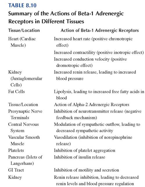
• β1 receptors: These receptors are primarily found in the heart, where they mediate the effects of sympathetic stimulation on cardiac function. Activation of beta-1 receptors leads to positive chronotropic effect and inotropic effect, and increased conduction velocity through the atrioventricular node (Table 8.10).
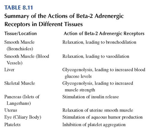
• β2 receptors: Found in smooth muscle cells, bronchioles of the lungs, walls of blood vessels, and the uterus, activation of these receptors leads to relaxation of smooth muscle, resulting in bronchodilation, vasodilation, and relaxation of the uterine muscle during pregnancy. Beta-2 receptors are also found in hepatocytes, where their activation leads to glycogenolysis and gluconeogenesis (Table 8.11).
• Beta-3 adrenergic receptors (β3 receptors): These receptors are found in adipose tissue and the urinary bladder. Their activation in adipocytes leads to lipolysis, while activation
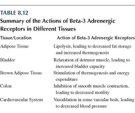
in the urinary bladder leads to relaxation of the detrusor muscle, promoting bladder filling (Table 8.12).
8.17