REPRODUCTIVE SYSTEM
Male
In adults the scrotum is easily visible ventrolateral to the anus. The skin of the scrotum is thin and covered with fine hairs. The inguinal canal remains open throughout life and has a distended diameter of 8-12 mm, which allows the testis to have a scrotal or inguinal position.
The testes descend between 30-40 days (Bivin et al. 1979; Greene 1962; Hebel & Stromberg 1986g).The testis is oval in shape and measures 20?14 mm. The head and tail of the epididymis are often sites of extremely large fat pads. The penis can easily be extruded from the prepuce and has an os penis (Hebel & Stromberg 1986g).
Accessory genital glands
Male rats have highly developed accessory sex glands (Bivin et al. 1979; Hebel & Stromberg 1986g) (Fig. 10.21). These produce a gelatinous copulatory plug, which is visible in the vagina post mating and is thought to prevent semen leaking out.
Seminal vesicles - These lie dorsolateral to the urinary bladder and are in contact with the rectum dorsally. They are large and lobulated, opening into the lower end of the vas deferens. Each is enclosed in a capsule together with the coagulating gland.
Prostate gland - This is bilateral and has three lobes: the dorsal, the lateral, and the ventral. The dorsal lobe, also known as the coagulating gland, lies close to the seminal vesicles (Komarek et al. 2000b).
Ampullary glands - These are the glands of the ductus deferens and lie near the bladder.
Bulbourethral glands - These are found near the urethra as it exits the pelvis.
Preputial glands
Preputial glands lie in the subcutaneous fat near the penis and open into the prepuce. These are sebaceous glands that secrete a pheromone used for scent marking. These regress with ageing and the glands can fill up with stagnant sebum. An analogue is also present in the female (Hebel & Stromberg 1986g; Komarek et al.
2000b).Female
The right ovary is located at the level of L4-5 just caudal to the right kidney. The left ovary lies at L5-6 caudal to the left kidney. The left ovary is nearer the midline than the right. Both are embedded in fat (Bivin et al. 1979; Hebel & Stromberg 1986g). The oviduct is convoluted and winds around the ovary in 10 to 12 garland-like loops. The uterus is duplex, meaning the two parts are separate along its entire length, uniting only at the vagina (Greene 1962; Hebel & Stromberg 1986g; King & Custance 1982). There is partial fusion caudally for 7-10 mm where they share the outer longitudinal layer of myometrium. Like most mammals an anastomosis between the ovarian artery (a branch of the aorta) and the uterine artery (a branch of the internal iliac) occurs in the uterine mesentery (Del Campo & Ginther 1972).
The mesovarium and mesometrium contain voluminous amounts of fat, which surrounds the kidneys, abdominal wall, and intestinal loops. There are no vaginal glands. In the female rat the urethra and vaginal orifice are completely separate. The only genital structure connected with the urinary system is the clitoris. The urethra lies cranially at the base of the clitoris and both are located in a high cone with clitoral glands. The vaginal opening lies caudal to this and is closed by a membrane until puberty in the female. The female has six pairs of mammary glands (Bivin et al. 1979; Hebel & Stromberg 1986g).
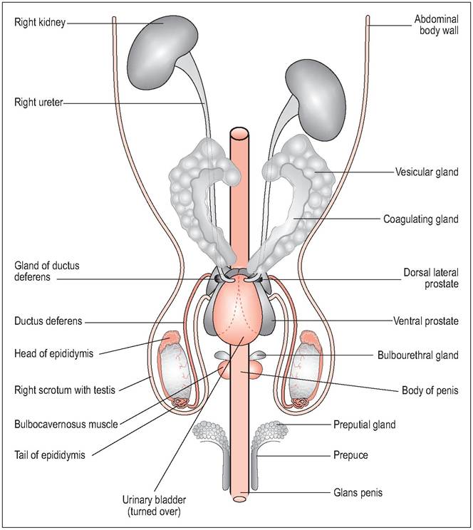
Figure 10.21 • Ventral view of male genital tract showing the extensive accessory glands.
Clitoral (preputial) glands
These paired glands lie in the subcutaneous fat near the clitoris and open into a preputial fold. These are sebaceous glands that secrete a pheromone used for scent marking (Komarek et al. 2000b).
Mammary glands
The glandular tissue changes volume and appearance during the estrous cycle, pregnancy, and lactation. Prior to the first pregnancy these glands consist of a few short tubules around the teats but these expand into each gland before parturition.
The cervical gland is rudimentary whereas the thoracic gland is diffuse, contains three nipples, and surrounds the base of the forelimb. The abdominal gland is only marginally developed while the inguinal gland has three nipples and surrounds the base of the hindlimbs (Komarek 2000). The thoracic and inguinal glands act as two discrete complexes, separated by a space just behind the ribs (Fig. 10.22). During lactation the thoracic glands expand rostrally as far as the parotid and mandibular glands, cranially and medially over the upper arm, and along the lateral thoracic wall. Caudally, the inguinal gland covers the lateral abdominal wall right back to the stifle and the anal region (Hebel & Stromberg 1986e; Komarek et al 2000b; Maeda et al. 2000).
At the base of each teat the transition zone between hairless and hair has large sebaceous glands. There are no teats in the male although mammary tissue can still be found in the corresponding area (Hebel & Stromberg 1986e).
Reproductive physiology
Photoperiod has a strong influence on the hypothalamus, which means it also affects the estrous cycle. The luteinizing hormone (LH) surge is strongly linked to the circadian rhythm and usually occurs in the late afternoon. Stress and suckling suppresses LH secretion and, hence, ovulation (Maeda et al. 2000).
Estrous cycle
Females are polyestrous, with a 4-5 day estrus. Ovulation occurs 9-10 hours after the commencement of estrus. Vaginal
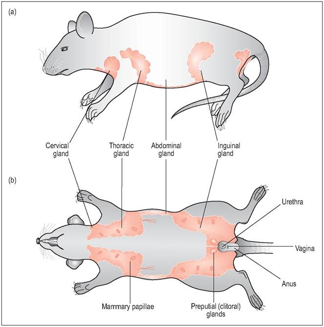
Figure 10.22 • Mammary tissue of the female rat is extensive, reaching from the neck and elbow to the inguinal region. There are six mammary glands on each side in the rat - three in the thorax, I abdominal, and two in the inguinal region.
(a) Lateral view of mammary glands
(b) Ventral view of mammary glands
From Popesko, P., Rajtova, V., & Horak, J. (1990) A colour atlas of anatomy of small laboratory animals.
Vol. 2. Aylesbury, UK: Wolfe with permission.CLINICAL NOTE
Mammary tumors are common in rats, and due to the extensive distribution of the mammary tissue, can appear anywhere from the axilla to the groin. They can occur in both males (16%) and females and incidence increases at over 18 months of age (Altman & Goodman 1979). About 80 to 90% are benign fibroadenomas (Percy & Barthold 2001) so surgical removal is recommended (Figs. 10.23 and 10.24). They can be associated frequently with pituitary tumors (Altman & Goodman 1979).
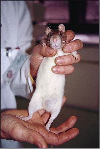
Figure 10.23 • Female rat with large tumor of inguinal mammary gland.
smears can be used to identify the phase of the cycle as estrogen causes a proliferation of vaginal epithelial cells during proestrus (Harkness & Wagner 1995) (Table 10.1).
Gestation
The gestation period is 21-23 days. The rat has a hemochorial placenta. There is little change in the uterus in the first trimester but after this it moves ventrally, displacing intestinal coils cranially. Parturition is usually by day and is quick, taking about 90 minutes to produce a litter which can vary in size from 3 to 18 (Maeda et al. 2000).
Birth
The pups weigh 5-6 g at birth and are altricial, with closed ears and eyes (Harkness & Wagner 1995). They have no intrinsic thermoregulatory mechanisms until the end of the first week of life so are kept warm by siblings and the mother (Fallon 1996; Koolhaas 1999). The ears open within 4 days and eyes open by the end of the 2nd week. Full hair has grown by the 7-10thday (Baker 1979; Fallon 1996; Koolhaas 1999).