Sense Organs
In vertebrates, including mammals, the major organs associated with detection of environmental stimuli—the eyes, ears, nose, and tongue—are concentrated in the head. Imbedded in these organs are cells of the nervous system specialized for sensing changes in external conditions.
The sensory information is transmitted to the appropriate areas of the brain where interpretation occurs, allowing sophisticated perception and appropriate action.TONGUE
The tongue in most mammals actually is employed in a variety of activities—feeding, drinking, grooming, as well as gustation (tasting). The receptors associated with gustation are located within papillae scattered over the surface of the tongue. Recall that the various papillae and their distribution were discussed in the digestive system [Figure 4-7B].
NOSE
The nose functions during respiration and olfaction (smelling). The sense receptors involved with detection of odoriferous stimuli are located within the epithelium of the mucous membranes lining the nasal cavities.
EAR
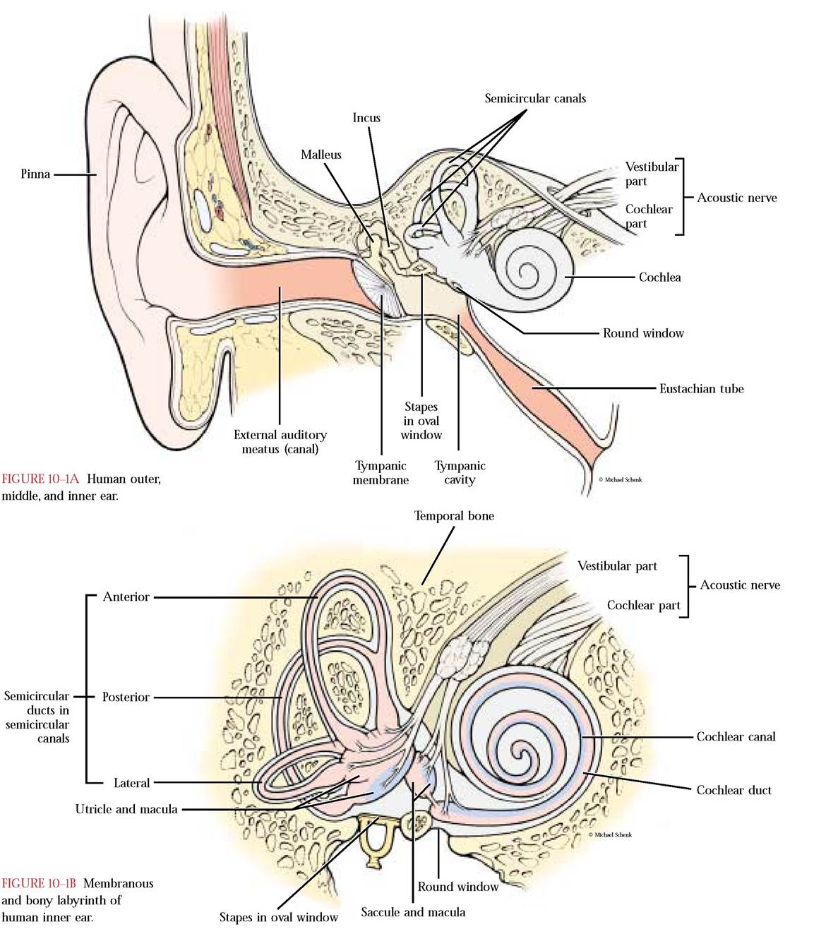
The ear of mammals is very difficult to dissect since it is imbedded within the dense petrous portion of the skull. It is best to study the anatomy of the ear using models. The ear consists of the external, middle, and inner regions. The external ear, also known as the pinna, is easily identifiable in most mammals and functions as a collecting funnel for sound waves. This sound energy passes through the external acoustic meatus and causes the tympanic membrane (eardrum) to vibrate. The vibration is transmitted sequentially to the inner ear through a series of three ear ossicles—the malleus, the incus, and the stapes—located in the tympanic cavity of the middle ear. The footplate of the stapes fits into the oval window, transmitting vibrations to the fluid filled cochlea in the inner ear.
The bony labyrinth is the cavity in the petrous bone in which rests the membranous labyrinth consisting of a blind tube (the cochlear duct), shaped like a snail shell, that is continuous with the organ of equilibrium consisting of the saccule, utricle, and three semicircular ducts.
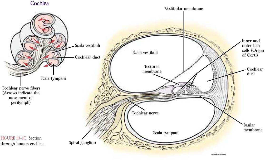
In the human, the semicircular ducts are identified as anterior, posterior and lateral. The inner ear rests in the bony labyrinth of the petromastoid portion of the skull.Within the membranous labyrinth circulates the endolymph while within the bony labyrinth circulates the perilymph. Located within the cochlear duct is the organ of Corti. Waves of fluid within the cochlea cause bending of the microvilli of the hair cells of the organ of Corti and produce nerve impulses that are transmitted by way of
cochlear nerves to the brain for sound interpretation. The saccule, utricle, and semicircular ducts are involved with equilibrium [Figure 10-1A, Figure 10-1B and Figure 10-1C].
EYE
External Structures
The eyeball of the cat is also difficult to dissect because one must remove it from the head, but an excellent substitute, a cow or sheep eye, is readily available. It is advantageous to dissect two eyes, one to study the extrinsic eye muscles that position the eyeball within the orbit and the other to view the internal eye structures.
To facilitate the study of the eye muscles, carefully remove the white connective tissue and fat, avoiding removal of any pink, tan or gray structures which may be muscles or glands from the eyeball. The optic nerve, which is white and located on the back and near the center of the eyeball, must be conserved [Figure 10-2A]. All mammals possess six extrinsic eye muscles—a ventral oblique, a dorsal oblique, a medial rectus, a lateral rectus, a dorsal rectus, and a ventral rectus. In order to identify these muscles correctly, it is imperative to determine whether the eyeball is left or right. The key to this determination is identification of the ventral oblique muscle. This muscle is found on the ventral surface (bottom) of the eyeball and is the only muscle that naturally wraps around the circumference of the eyeball.
Its insertion is on the lateral aspect of the eyeball very near the insertion of the lateral rectus muscle. The cut end of the ventral oblique muscle represents the medial aspect of the eyeball [Figure 10-2A]. You now should be able to identify the dorsal, ventral, medial, and lateral aspects of the eyeball. Identify the ventral rectus muscle over which the ventral oblique muscle lies. Next identify the medial rectus muscle appearing approximately opposite the lateral rectus muscle. Continue around the eyeball dorsally and identify the dorsal rectus muscle that inserts approximately opposite the ventral rectus muscle. Finally, identify the dorsal oblique muscle whose insertion and muscle direction is as its name implies, somewhat oblique, and opposes the ventral oblique muscle [Figure 10-2A].In the cat, but not the human, on the posterior surface of the eyeball and surrounding the projecting optic nerve, is a four-part muscle, the retractor bulbi [Figure 10-2A]. This muscle retracts the eyeball. Another muscle, the levator palpebrae superioris, attached to the upper eyelid and found on the anterio-dorsal surface of the eyeball, lifts the eyelid. You may see yet another muscle, the orbicularis oculi, that is arranged in a circular sphincter-like pattern in and around the eyelids. It functions during forced closure of the eyelids, e.g., when the cat is being disciplined. If the eyelids are missing, these muscles will not be observed.
Eyeball moisture for most terrestrial vertebrates affords a well lubricated surface to assure that the surface remains
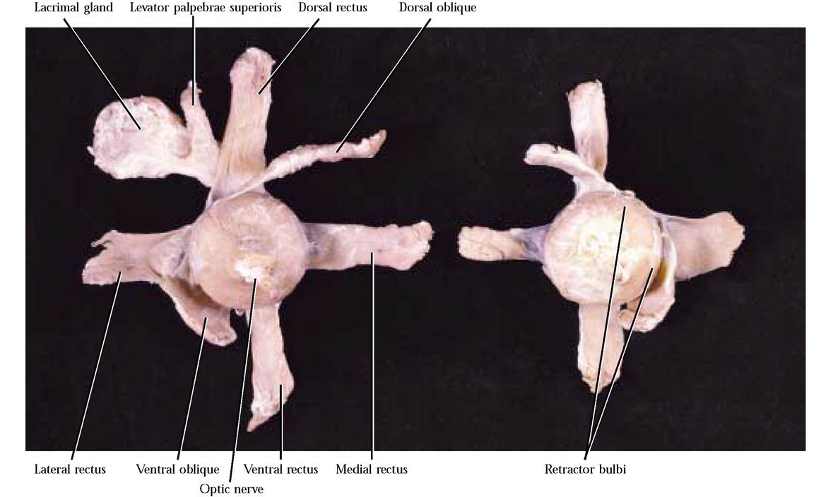
FIGURE 10-2A Extrinsic eye muscles; left eye and right eye.
clean and can function as part of the lens system. To provide this moisture, three glands are present. A lacrimal gland, also present in humans, located on the dorsolateral aspect of the eyeball, secretes a solution known as tears [Figure 10-2A].
This secretion moves across the eyeball, keeping it moist, and then draining into the nasolacrimal duct and eventually into the nasopharynx. A small salivary gland, the infraorbital gland, not found in humans, is located in the orbit beneath the eye. It drains into the mouth. Within a transparent fold, the nictitating membrane [Figure 10-2B], located in the medial angle of the eye, the small harderian gland may be identifiable. Its secretions, like that of the lacrimal gland, bathe the eyeball. This gland is not present in humans. The nictitating membrane acts as a protective mechanism in many mammals, but is vestigial in humans.Internal Structures
If you received two eyeballs, dissect the one not used for external structures. When only one eyeball is available, use caution during the following dissection so that you preserve muscles, etc., previously identified.
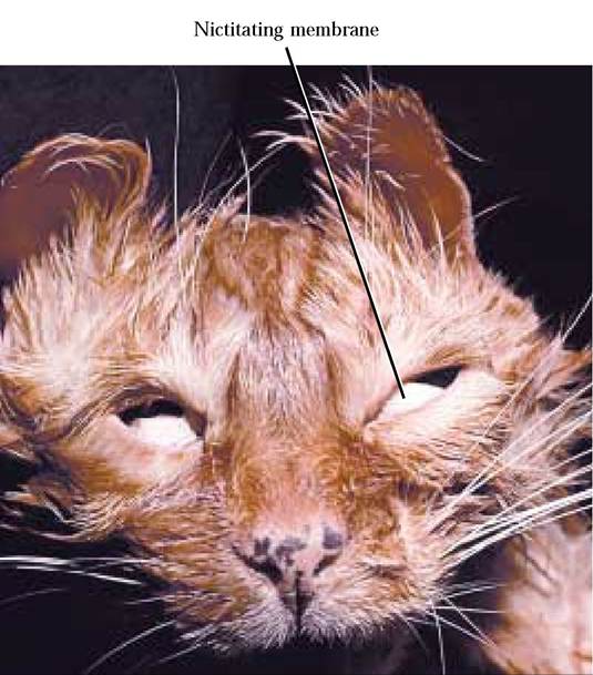
FIGURE 10-2B Nictitating membrane.
To prepare a specimen that is superior for the study of internal structures, insert the tip of a sharp scalpel into the eyeball approximately one centimeter from the posterior edge of the cornea and continue to cut around the circumference of the eyeball to separate it into anterior and posterior parts. Store the anterior and posterior parts of the eyeball in the open air at room temperature for 24-48 hours or until the jelly-like substance has dried out. This gelatinous substance, the vitreous humor, occurs between the lens and the retina and functions in holding the retina and lens in place and serves as a refractive medium as part of the lens system of the eye. Between the cornea and the ciliary body is the aqueous humor, a fluid continuously secreted by the ciliary body into this space. The aqueous humor is also continuously removed through a venous sinus surrounding the eye, therefore under normal conditions its volume remains constant. It functions in keeping the structures of the eye in position as well as serving as part of the lens system of the eye.
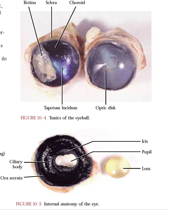
Further, it supplies the structures in this area with area, the tapetum lucidum, is obvious [Figure 10-4]. This is common in vertebrates that are active in subdued light. Have you noticed that cat eyes when suddenly illuminated by incidental light in the dark appear yellow or green? It is light reflecting from the surface of their tapetum lucidum. Gently move the retina to one side to see its point of attachment at the back of the eyeball. This point of attachment is the optic disk [Figure 10-4]. Through this area, pass the neurons of the optic nerve and nutrient blood vessels. The optic disk is the area known as the blind spot since there are no photoreceptors, rods, and cones, located there.Anteriorly, the choroid is modified as the iris. It consists of smooth muscle arranged radially and circularly around an opening known as the pupil and regulates its diameter dependent upon incident light [Figure 10-3 and Figure 10-5]. Pigment of the iris influences eye color. The iris is continuous with a second modification of the choroid, the ciliary body. The ciliary body consists of ciliary muscles and the ciliary process. The delicate zonule fibers of the
nutrients and oxygen while removing metabolites.
Three distinct concentric tunics or layers form the eyeball. In the posterior end of your preparation, identify the three layers. The outer layer is a tough white, fibrous coa the opaque sclera. Over the anterior surface of the eyeball the sclera is modified as a transparent window called the cornea [Figure 10—3]. The middle layer, the choroid, is heavily pigmented and appears black. The retina, the inne most tunic, consists of an outer pigmented layer abutting the choroid and an inner neural layer abutting the vitreou humor. The neural layer will appear creamy, folded and displaced from its normal position due to preservation wh the pigmented layer generally associates with the choroid. Because of this displacement, an iridescent bluish-green suspensory ligament extend from the ciliary body to the lens.
The ciliary muscles are smooth muscles that regulate lens shape. The folded ciliary process secretes the aqueous humor. The scalloped junction along the posterior aspect of the ciliary body is known as the ora serrata [Figure 10—5]. It marks the anterior margin of the neural portion of the retina.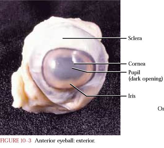
The lens is a biconvex transparent structure that is responsible for fine focusing [Figure 10-6]. In your specimen, the lens will appear opaque, due to the preservation process.
The anatomy of the human eye is virtually identical to that of the cat. Figure 10-7 represents a cross section of a human eye.
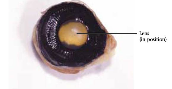
FIGURE 10-6 Lens in natural position.
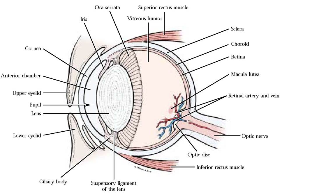
FIGURE 10-7 Sagittal section of human eye.