Nervous System
The nervous system is one of the most complex and least understood of any of the systems in animals. The complexity of this system is reflected not only in the anatomy but also in its physiology.
Consider that this system, often in conjunction with portions of the endocrine system, is the controlling center for the homeostatic well-being of the body. These systems initiate, moderate, and coordinate all body activities.Characteristic of the most complex of any known animal nervous system, that of vertebrates, is a dorsal hollow nerve cord with a greatly elaborated anterior end known as a brain. Invertebrate animals are often said to possess a brain but it consists of a mass of nervous tissue called ganglia and is generally capable of behavior associated with stereotypic activities.
Originating from the vertebrate brain are 10-12 pairs of cranial nerves that generally innervate glands, muscles, and organs at the anterior end of the animal, with the exception of the vagus nerve that innervates most of the visceral organs. Cranial nerves are associated with various regions of the brain and pass through specific foramina of the vertebrate skull.
The dorsal hollow nerve cord, mentioned above, is more often referred to as the spinal cord. It extends from the brain, passes through the foramen magnum, and continues through the vertebral foramen of the spinal column made up of vertebrae. Paired spinal nerves, associated with the innervation of skin and its structures and muscles of individual body segments, emerge between individual vertebrae.
Among tetrapods, with their more sophisticated appendage movement, a more complex arrangement of the simple primitive segmental spinal nerve pattern is encountered in the forelimb and hindlimb region of the spinal cord. In the forelimb region, this arrangement is known as the brachial plexus while in the hindlimb region the arrangement is called the lumbosacral plexus.
Classically, the brain and spinal cord are grouped together as the central nervous system while the cranial nerves, spinal nerves, and autonomic nerves are known as the peripheral nervous system.
THE BRAIN
The mammalian brain, in many ways, is unique among vertebrates. The most prominent portions are the cerebrum and cerebellum, both of which have evolved in a similar fashion to accommodate the immense increase in neurons and supporting cells of this part of the central nervous system. The surfaces of both exhibit numerous convolutions (gyri) and crevices (sulci) as a means to substantially increase the surface area of the cerebrum and cerebellum with the result that many more cells can be fit into these areas while still conserving space in the cranium. In many instances in the cerebrum, the gyri and the sulci each have a specific name. More than likely, mammal heads would be even larger than they are without this design. Further, in contrast to nonmammalian vertebrates, the cerebrum and cerebellum of mammals possess a thin outer layer of gray matter, the cortex, consisting mainly of nerve cell bodies and unmyelinated nerve fibers overlying the main mass of white matter consisting primarily of myelinated
nerve fibers. The prominent paired optic lobes of the mesencephalon, the major integration and coordination center of nonmammalian vertebrates, appear in mammals as two paired lobes, the corpora quadrigemina, now a center for the regulation of auditory, visual and other reflex activities since the original functions now reside in the cerebrum and cerebellum.
Three primitive embryological divisions of the brain are recognized—the anterior prosencephalon, or forebrain, and the posterior rhombencephalon, or hindbrain, separated by the mesencephalon or midbrain. As differentiation occurs, the mesencephalon remains intact but the prosencephalon subdivides into the telencephalon and the diencephalon while the rhombencephalon divides into the metencephalon and myelencephalon, yielding a five-part brain.
Later events lead to the development of the olfactory bulbs and cerebrum from the telencephalon, the corpora quadrigemina from the roof of the mesencephalon, the cerebellum and pons from the metencephalon, and the medulla oblongata from the myelencephalon.Since accessing the intact brain of the cat is most difficult and often results in a poor specimen, we suggest using a sheep brain as representational of the typical mammalian pattern of anatomy. We have found that brains with intact cranial nerves and pituitary glands and with the meninges removed are the best dissection specimens. We also recommend that your instructor, using a sharp kitchen knife, cut each brain into two equal halves so that the internal sagittal anatomy can be studied.
THE MENINGES AND VENTRICLES OF THE BRAIN
The central nervous system is isolated from the rest of the body by three protective layers known as meninges. The innermost layer, the pia mater, remains in intimate contact with the brain surface and can be recognized by its abundant vascularity. The outermost whitish, tough fibrous layer, the dura mater, often remains adherent to the area surrounding the pituitary gland (hypophysis) even after removal of the meninges. The middle layer, the arachnoid membrane, is delicate, web-like and extends between the pia and dura maters.
Since the brain represents a phenomenal specialization of the anterior portion of the dorsal hollow nerve cord, it should not be surprising to learn that the brain possesses cavities known as ventricles. Ventricles I and II lie in the left and right cerebral hemispheres, respectively. They are each connected by a small canal to ventricle III that occurs in the diencephalon which is, in turn, connected by a canal called the cerebral aqueduct in the mesencephalon to ventricle IV, lying in a V-shaped space of the medulla oblongata. Each of these ventricles is roofed by a complex of tissues called a choroid plexus. A choroid plexus consists of the inner lining of the brain, the ependyma, and a highly vascularized portion of the pia mater.
This is the site of the “blood-brain barrier ” that is effective in preventing the contamination of the central nervous system by such potentially damaging invaders such as bacteria, viruses, etc. A specialized secretion, the cerebrospinal fluid, is produced by the choroid plexi. Cerebrospinal fluid circulates within the ventricles of the brain, the central canal of the spinal cord and in the subarachnoid space between the pia mater and arachnoid membrane in mammals. It functions as a lubricant, prevents damage from mechanical shock, and also acts as a buoyant fluid, allowing the brain to float and be perceived as a fraction of its actual weight.EXTERNAL ANATOMY
The Telencephalon
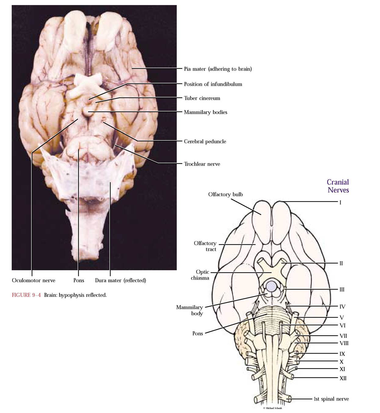
From the dorsal view, observe the cerebrum consisting of paired cerebral hemispheres separated from each other by the deep longitudinal cerebral fissure. Notice the numerous gyri, elevations, and sulci, shallow depressions, sculpting the surface [Figure 9-1]. On the ventromedial aspect of the cerebrum lie the olfactory bulbs. The bulbs lie above the cribriform plate of the ethmoid bone and receive fibers from neurons in the olfactory epithelia lining the nasal cavity. Those fibers represent part of the olfactory nerve (I) that carry sensory information to the cerebrum. Two flat bands, the more conspicuous lateral olfactory band and the medial olfactory band lead from the olfactory bulbs toward the olfactory portion of the brain, the pyriform lobe, that is separated from the rest of the cerebrum by a somewhat indistinct rhinal sulcus. A triangular area between the two bands is often recognized as the olfactory trigone [Figure 9-2].
With two exceptions, the human cerebrum is very similar. In addition to being considerably larger, the human brain is more complexly folded and subdivided into recognizable lobes and the olfactory bulbs are smaller.
The Diencephalon
Since the cerebrum has developed to such an extraordinary degree in mammals and has attained a size that brings it into contact with the second largest portion of the brain, the cerebellum, a great deal of the diencephalon can be best seen in the sagittal section and in the dissection of the brainstem to be done later.
Three areas, the floor or hypothalamus, the sides or thalamus, and the thin roof, the epithalmus, enclose the third ventricle of the diencephalon. A major landmark, the optic chiasma, an X-shaped structure, demarcates the cranial end of the hypothalamus on the ventral surface of the brain. The two stout bands of tissue at the cranial end of the chiasma are the optic nerves (II) that consist of nerve fibers carrying sensory information from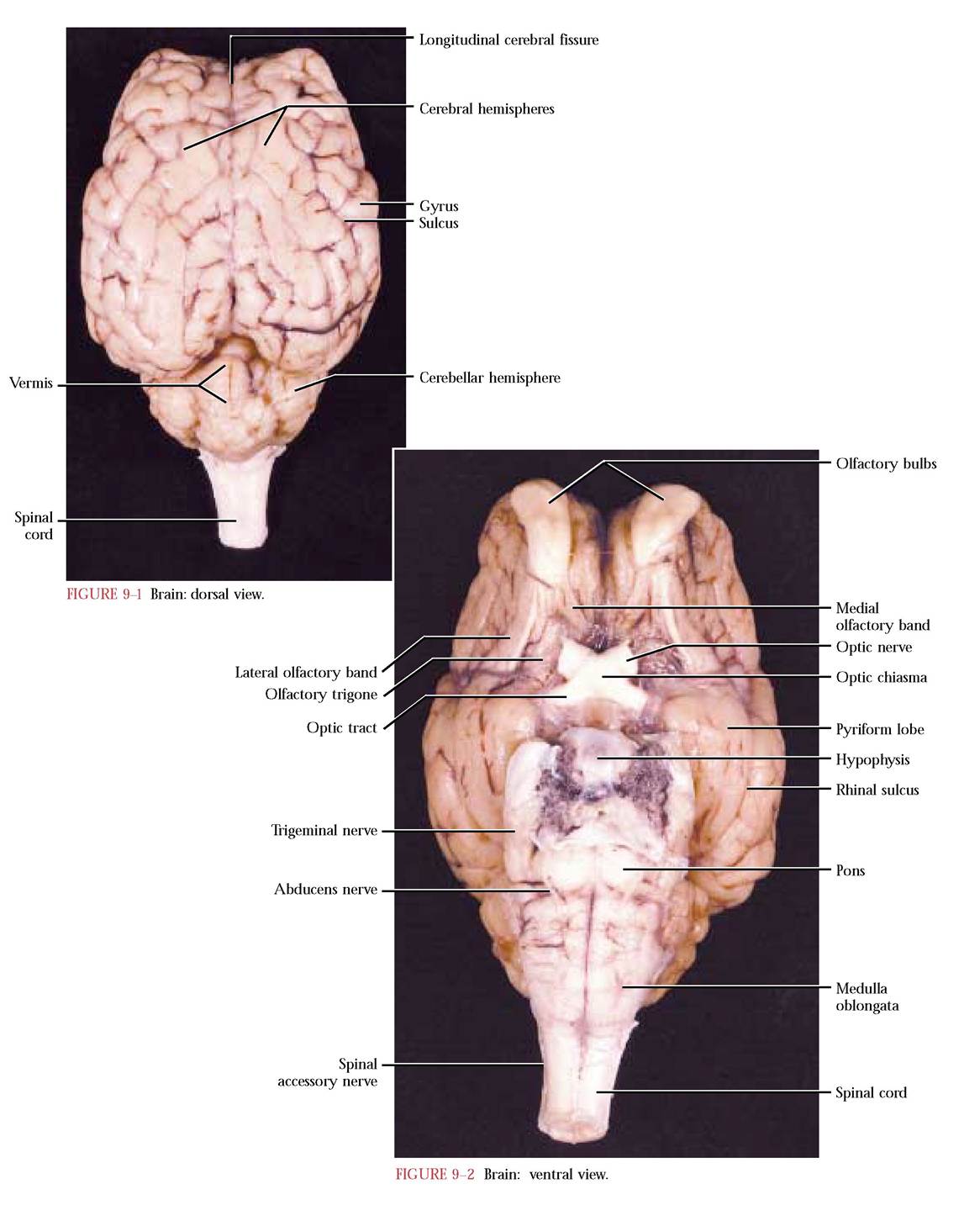
the eyes. At the chiasma, some fibers from each eye cross over to the opposite side while others pass straight through, carried via optic tracts to their respective sides [Figure 9-2, Figure 9-3A, and Figure 9-3B]. This circuitry allows mammals to see life three dimensionally. A delicate slender tube, the infundibulum, extends from the hypothalamus to the pituitary gland (hypophysis). The slightly convex area of the hypothalamus just posterior to the infundibulum is the tuber cinereum. Just caudal to the tuber cinereum are the paired mammilary bodies, marking the caudal end of the hypothalamus [Figure 9-3A and Figure 9-3B].
The Mesencephalon
While gently spreading the cerebrum and cerebellum apart, observe the bulges representing the corpora quadrigemina. These bodies consist of a larger, more prominent, pair of superior colliculi resting on a smaller less conspicuous pair of inferior colliculi. On the ventral surface observe elongated paired longitudinal cerebral peduncles representing bundles of nerve fibers extending between anterior and posterior areas of the brain [Figure 9-3A and Figure 9-3B]. The yellowish flat bands emanating from the ventral surface of the cerebral peduncles and often adhering to the dura mater attached in the region of the hypophysis are the oculomotor nerves (III). The trochlear nerve (IV) is unique because it is the only cranial nerve to emerge from the dorsal surface of the brain, the mesencephalon just anterior to the pons [Figure 9-4].
The Metencephalon
This region of the brain consists of a ventral bulging band of transverse fibers, the pons, located just posterior to the cerebral peduncles. The largest and most conspicuous of the cranial nerves, the trigeminal (V), emanates from the most posterior region of the pons. By far, the larger portion of the metencephalon consists of the dorsal cerebellum. Notice the medial vermis, flanked by paired cerebellar hemispheres [Figure 9-1, Figure 9-2, and Figure 9-3B].
The Myelencephalon
The medulla oblongata contains fiber tracts permitting communication between the brain and the spinal cord [Figure 9-2, Figure 9-3A, and Figure 9-3B]. Cranial nerves VI, the abducens; VII, the facial; VIII, the acoustic; IX, the glossopharyngeal; X, the vagus; XI, the spinal accessory; and XII, the hypoglossal, are all associated with the medulla oblongata. The spinal accessory, as its name implies, also receives fibers from the anterior end of the spinal cord [Figure 9-5].
In the human brain, the most obvious difference is size and less obvious is its shape. The brain is larger and less elongate with the cerebellum and medulla tucked under the immense cerebrum. Have you ever wondered why your sense of smell is not as good as your dog or cat? There are two reasons: the area of human olfactory epithelium is a fraction of that of other mammals and the olfactory bulbs are greatly reduced.
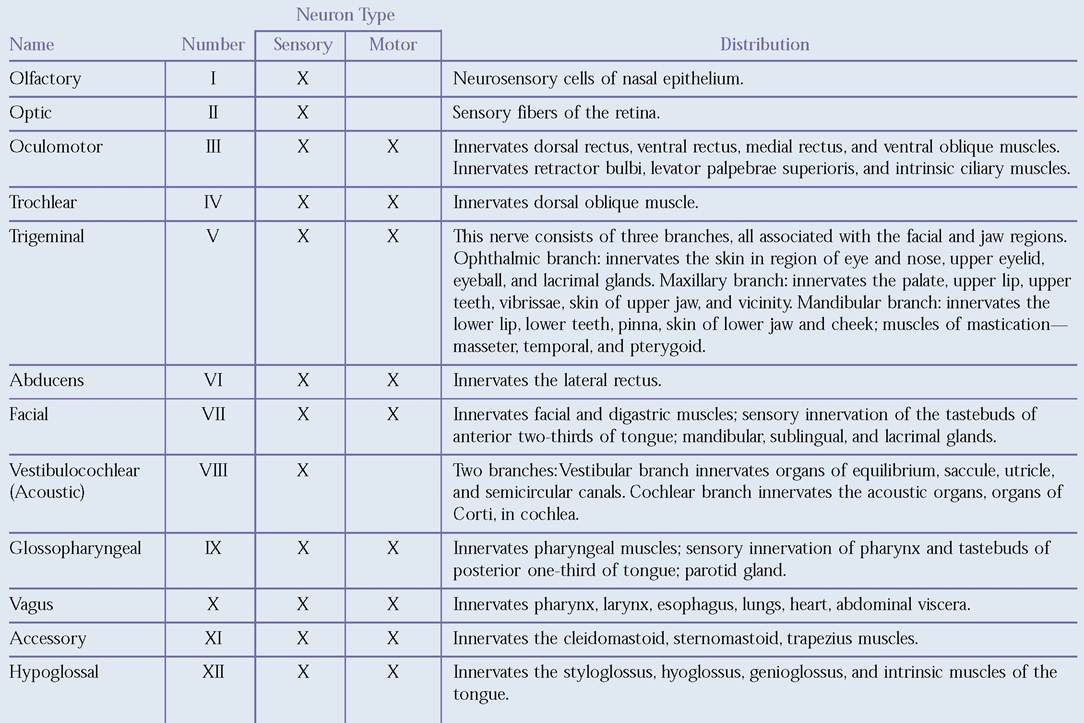
Table of Cranial Nerves
Internal Anatomy—Sagittal Section of the Brain
A careful sagittal section usually results in two halves that are very similar. External features just discussed, in many cases, will be seen in this dissection, sometimes in greater detail. It is easier to see the relationship of the cerebrum to the diencephalon and mesencephalon. The thin layer of gray matter (actually tan in preserved specimens) on the outer surface of the cerebrum and cerebellum is very obvious in this section. In the following discussion, refer to Figure 9-3A, Figure 9-3B, and Figure 9-6].
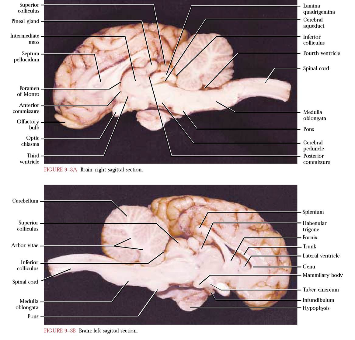
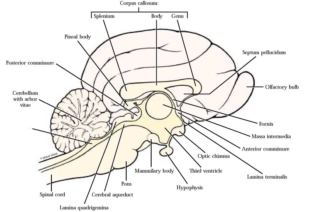
Within the three groups of mammals living today, the monotremes or egg-laying mammals, the marsupials, and the placental mammals, it is only among the placental mammals that a corpus callosum has evolved. It consists of transverse nerve fibers that permit transmission of nerve impulses between the cerebral hemispheres. The cranial curve of the corpus callosum is known as the genu and the caudal end is called the splenium, with the trunk extending between them. Another paired bundle of fibers, the fornix, more extensive than seen in this section, appears ventral to the corpus callosum. Connecting the corpus callosum and the fornix is the thin wall of tissue called the septum pellucidum. It consists of a double sheet of gray and white matter that separates the two lateral ventricles of the cerebrum. Very often, the sagittal cut will have left the septum entirely in one of the brain halves. If that is true, it will be easy for you to observe one of the lateral ventricles, on the side devoid of the septum.
Now, direct your attention to the area almost directly ventral to the regions just discussed. If the sagittal cut was successful in approximating the midsagittal plane, a rather prominent circular mass, the intermediate mass, will be distinguishable residing within a shallow irregular space, the third ventricle and cerebral aqueduct. Each of the cerebral hemispheres contains a lateral ventricle that communicates with the third ventricle by way of the foramen of Monro. Recall that the third ventricle is enclosed within the walls of the diencephalon. The aqueduct sits within the confines of the mesencephalon leading to the fourth ventricle resting within the medulla oblongata. The intermediate mass is a central connective bridge between the left and right thalamus that make up the two lateral walls of the diencephalon.
Fourth ventricle
Actually, all of the tissue lateral to the third ventricle in this section, with the exception of the single layered ependymal lining the ventricle, is thalamus. The floor and ventral walls of the diencephalon consists of the hypothalamus. Observe the relationships of the optic chiasma, infundibulum, hypophysis, and mammilary bodies, all associated with the hypothalamus and seen in external view. The thin lamina terminalis forms the cranial wall of the diencephalon. Notice the small thickening, the anterior commissure, in the dorsal portion of the lamina terminalis. The epithalamus is the very thin roof of the diencephalon. The most prominent structure here is the pineal gland or body projecting from its surface. Just cranial to it is the habenular trigone and ventral to it is the posterior commissure.
Features of the mesencephalon include the dorsally located corpora quadrigemina, consisting of a pair of superior colliculi, controlling visual reflexes, and a pair of inferior colliculi, controlling auditory reflexes and resting on the lamina quadrigemina. The thick floor consists of the cerebral peduncles, bundles of nerve fibers extending between the cerebrum and other brain areas. The usually narrow space enclosed in the mesencephalon is the cerebral aqueduct connecting the third and fourth ventricles.
The rounded protuberance posterior to the cerebral peduncles is the pons and the somewhat elongated, flattened region just caudal to the pons is the medulla oblongata. Caudal to the medulla oblongata the central nervous system continues as the spinal cord whose cavity is the central canal.
The large, deeply grooved body lying dorsally over the fourth ventricle and pons and medulla oblongata is the cerebellum. Note the similarity in tissue arrangement between the cerebrum and cerebellum, with the outer layer of gray matter lying over the white matter. In sagittal section, this relationship of the tissues of the cerebellum suggested a treelike construction to early anatomists and they named this arrangement of white matter in the cerebellum the arbor vitae (tree of life).
The Brainstem
The brainstem of mammals consists of the mesencephalon, pons, and medulla oblongata. It is the site of a number of relay and vital reflex centers. The specimen in Figure 9-7A includes portions of the diencephalon, as well. To produce a dissection similar to Figure 9-7A grasp the cerebrum of one of your sagittal sections and carefully separate it from the rest of the section. Now slice through the thalamus of the diencephalon to create a specimen similar to Figure 9-7A. Carefully lift up the cerebellum and remove it from the brainstem using a sharp knife or scalpel, while watching its connection to the underlying tissue so that tears do not occur in the area. After removal, if necessary, use a sharp scalpel to make a smooth slice through the anterior, middle, and posterior cerebellar peduncles similar to Figure 9-7A.
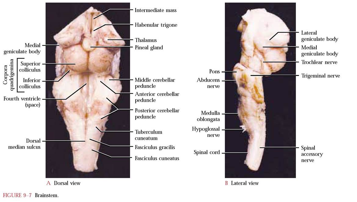
Beginning with structures associated with the diencephalon, not part of the brainstem, locate the thalamus, intermediate mass, the habenular trigone, and pineal gland. Notice the two subtle bulges on the lateral wall of the thalamus. The more anterior and less obvious of these diencephalic structures, the lateral geniculate body, associated with the superior colliculus, part of the mesencephalon and anterior portion of the brainstem, is involved in visual function, while the posterior and more obvious medial geniculate body, associated with the inferior colliculus, also part of the brainstem, serves as a relay of auditory impulses [Figure 9-7A and Figure 9-7B].
Locate the anterior, middle, and posterior cerebellar peduncles, representing bundles of fibers connecting the cerebellum to the brainstem. The space existing between the Y-shaped halves of the medulla oblongata is the fourth ventricle. This Y-shaped configuration consists of fiber tracts transmitting sensory impulses between the spinal cord and the medulla oblongata. Note the shallow central dorsal groove, the dorsal median sulcus. The two halves of the Y consist of the fasciculus gracilis. Lateral to the fasiculus gracilis, locate the fasciculus cuneatus and the tuberculum cuneatum [Figure 9-7A].
the arrangement in the cerebrum and cerebellum [Figure 9-8]. This portion of the central nervous system is the site of interconnecting synapses between sensory, association, and motor neurons involved in various reflex actions. Recall that the nerve cord in vertebrates is defined as dorsal and hollow. Examine the cut end of the spinal cord of the brain specimen and note the small central canal that constitutes the “hollow” part of the definition. The central canal is continuous with the fourth ventricle therefore the cerebrospinal fluid not only circulates within the brain ventricles, but also within the central canal of the spinal cord. Passage of the fluid through foramina in the fourth ventricle into the subarachnoid space allows circulation of the cerebrospinal fluid, thereby bathing the outer surface of the central nervous system.
In the body, the spinal cord lies within the vertebral canal formed by the articulated vertebrae. The spinal cord is not uniform in diameter, but exhibits two conspicuous enlargements, the anterior cervical that supplies nerves to the forelimb and the posterior lumbosacral that supplies nerves to the hindlimb. It continues posteriorly, ending in the tail as a slender thread of tissue called the filum terminalis. The human condition is very similar.
SPINAL CORD
SPINAL NERVES
Note that the more or less cylindrical spinal cord is continuous with and posterior to the medulla oblongata [Figure 9-3A, and Figure 9-3B]. Like the brainstem, the white matter in the spinal cord surrounds the gray matter, in contrast to There are 38-39 pairs of spinal nerves in the cat, 8 cervical, 13 thoracic, 7 lumbar, 3 sacral, and 7 or 8 caudal nerves.
Unlike the cat, the human possesses 31 pairs of spinal nerves, 8 cervical, 12 thoracic, 5 lumbar, 5 sacral, and 1 caudal
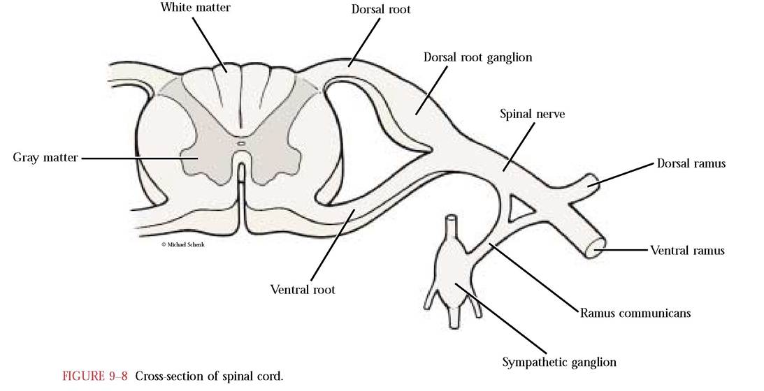
(coccygeal). Each spinal nerve generally carries both sensory and motor fibers. Spinal nerves are constructed at their spinal cord ends with a dorsal root carrying the sensory neurons and a ventral root carrying the motor neurons. They combine to form the spinal nerve, emerge between adjacent vertebrae as paired structures, and then subdivide into dorsal and ventral rami (branches) supplying their respective body segments [Figure 9-8].
Complex interrelationships of the ventral rami in certain body regions are identified as a plexus. Commonly, three of these, the cervical in the neck region, the brachial in the shoulder and forelimb region, and the lumbosacral in the hip and region of the hindlimb are discussed. In our treatment, the cervical and brachial are combined and discussed as the brachial plexus. In other treatments, spinal nerves 1-5 (cervical nerves 1-5) are referred to as the cervical plexus. Cervical nerves 6-8 along with the first thoracic nerve are referred to as the brachial plexus.
BRACHIAL PLEXUS
During the dissection of the muscular and circulatory systems, many of the nerves of this plexus were exposed. To do an orderly dissection of this complex, you will begin at its anterior end and work posteriorly. For the following dissection and discussion, see Figure 9-9. As reference points, locate the hypoglossal nerve (XII) which overlies the hyoglossus muscle previously discussed during the dissection of the neck muscles and the spinal accessory nerve (XI). To expose the spinal accessory nerve, carefully cut the sterno- mastoid and cleidomastoid muscles at their insertion ends. Because these muscles are innervated by this nerve, exercise great caution. Find a pair of swellings, the superior cervical and nodose ganglia, which occur just posterior to the hypoglossal nerve. Find the vagus nerve (X) extending posteriorly from the nodose ganglion and running along the common carotid artery in a sheath closely applied to the trachea. Although the vagus, spinal accessory, and hypoglossal are cranial nerves, they are important as references to a complete and orderly dissection. The nerves of the brachial plexus often have more than one point of origin, e.g., the first subscapular nerve arises from the sixth and seventh cervical nerves. In a plexus, interconnecting nerves carry fibers of the ventral rami from one spinal nerve to another, thereby contributing to the multiple origin of certain nerves in these complexes.
The first cervical nerve, quite small and difficult to recognize because of copious amounts of connective tissue in the area, passes medially over the ventral surface of the longus coli m. to supply ventral muscles of the neck.
Locate the second, third, fourth, and fifth cervical nerves, somewhat obscured by connective tissue and supplying the tissues of the shoulder and neck area.
Fibers of the fifth and sixth cervical nerves contribute to the formation of the phrenic nerve that travels lateral to and in close association with the vagus nerve through the thoracic region and innervates the diaphragm.
The sixth cervical nerve extends to the area of the shoulder joint where the prominent suprascapular nerve originates and passes with the transverse blood vessels between the supraspinatus and subscapularis muscles, supplying the supraspinatus and infraspinatus muscles. Notice that a small branch to the skin of the shoulder originates at the same point as the suprascapular nerve.
The sixth and seventh cervical nerves give rise to the first subscapular nerve that innervates the subscapularis m. and travels with the subscapular vessels. The axillary nerve originates from these two cervical nerves and travels with the posterior humeral circumflex vessels beneath the posterior edge of the biceps brachii m. to innervate lateral shoulder muscles such as the teres major, teres minor, deltoids, and cleidobrachialis. Likewise, the musculocutaneous nerve originates from the sixth and seventh cervical nerves and branches near the biceps brachii, innervating the biceps brachii, brachialis and coracobrachialis muscles as well as the skin of the forelimb.
The second subscapular nerve originates from the seventh cervical nerve and innervates the teres major m.
The anterior ventral thoracic nerve arises from the seventh cervical nerve, travels with the ventral thoracic vessels, and innervates the pectoral muscles.
The largest nerve in the plexus is the radial that originates from cervical nerves six, seven, eight and the first thoracic nerve, travels with the deep brachial vessels, and innervates the triceps brachii, epitroclearis, and extensor muscles of the forelimb.
The median nerve arises from cervical nerves seven, eight, and the first thoracic nerve, and travels with the brachial artery passing through the supracondyloid foramen to innervate some of the flexor muscles of the forelimb.
The posterior ventral thoracic nerve originates from the eighth cervical and first thoracic nerves, travels with the long thoracic blood vessels, and innervates the pectoral muscles.
The ulnar nerve arises from the eighth cervical and first thoracic nerves, travels parallel with the median nerve, crosses the medial epicondyle of the humerus, and innervates flexor muscles of the forelimb.
The medial cutaneous nerve originates from the first thoracic nerve and innervates the skin of the forelimb.
The third subscapular nerve originates from cervical nerves seven and eight, travels with the thoracodorsal vessels, and innervates the latissimus dorsi m.
The long thoracic nerve, lying along the lateral surface of and supplying the serratus ventralis m., originates from the seventh cervical nerve.
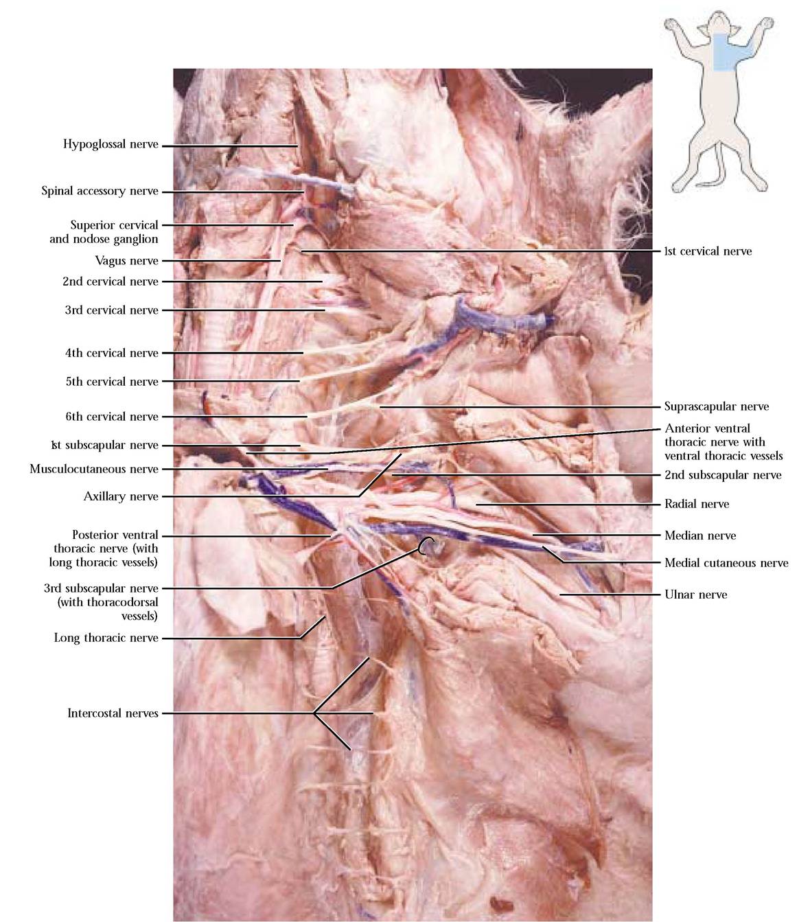
With the exception of the first thoracic nerve, the remainder of the thoracic nerves are referred to as the intercostal nerves, supplying intercostal muscles, lateral thoracic muscles, some abdominal muscles, back muscles, and skin.
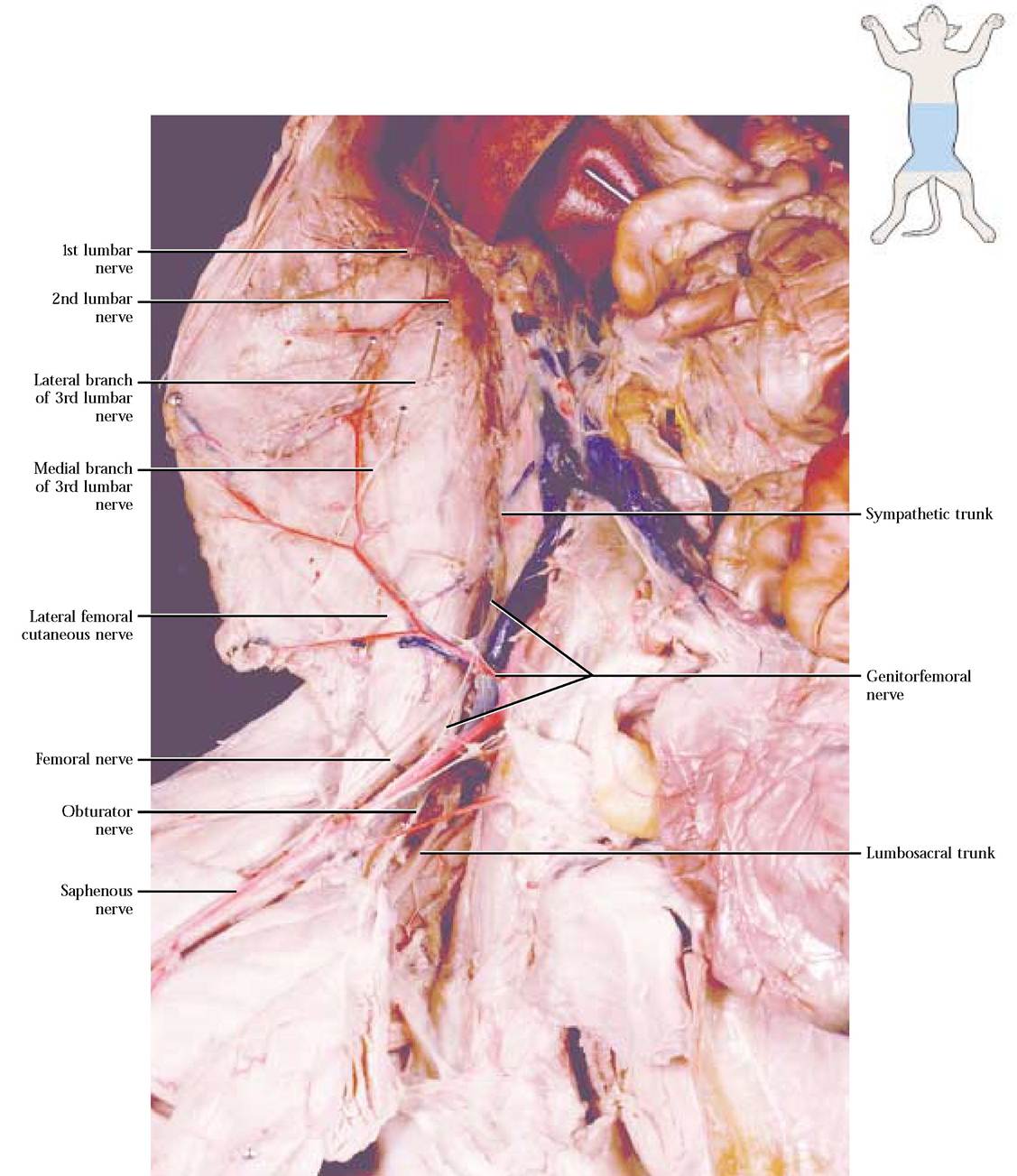
LUMBOSACRAL PLEXUS (VENTRAL VIEW)
The nerves that supply the lumbar and sacral regions and the hindlimb consist of a series of paired nerves, seven lumbar and three sacral, passing between adjacent lumbar and sacral vertebrae. Not surprisingly, the basic construction of nerves in this area mirrors those in the brachial region, in that they possess dorsal and ventral rami. The dorsal rami supply dorsal muscles and structures of the skin. The anatomical architecture of the ventral rami of the first three lumbar nerves consists of a medial and lateral branch that innervate muscles and structures in this region. The basic structural plan of the lumbosacral plexus consists of the ventral rami of lumbar nerves 4-7, sacral nerves 1-3, and communicating branches between these regions.
To study the architecture of the neural anatomy of these areas, we will begin by finding the medial branch of the third lumbar nerve as it emerges from beneath the illiopsoas m. and passes lateral to it. Trace it anteriorly to its source by carefully removing the anterior portion of the illiopsoas m. Care must be exercised in its removal since other lumbar nerves lie beneath it. Find the lateral branch of the third lumbar nerve. By removing the illiopsoas muscle, you should be able to find the second and first lumbar nerves with their medial and lateral branches [Figure 9-10A and Figure 9-10B].
The remainder of the dissection involves the lumbosacral plexus. One of the distinctive, but delicate and fragile nerves of this complex is the genitofemoral nerve that originates as the medial branch of the fourth lumbar nerve. An inconspicuous lateral branch of the fourth joins the fifth lumbar nerve. First, locate the genitofemoral nerve on the surface of the external iliac blood vessels [Figure 9-10A and Figure 9-10B]. Follow it anteriorly as it passes beneath the dorsal aorta and posterior vena cava to its source. In good dissections, particularly in males, you should be able to distinguish a genital branch innervating structures in the pelvic region.
The lateral femoral cutaneous nerve originates primarily from the fifth lumbar nerve with minor contributions from the fourth lumbar nerve [Figure 9-10A and Figure 9-10B]. It emerges from beneath the psoas minor m., passes over the illiopsoas m., travels with the deep ilial circumflex vessels, and supplies the lateral surfaces of the hip and thigh regions.
The majority of fibers of lumbar nerve six, in company with a branch of the fifth, form the prominent femoral nerve [Figure 9-10A and Figure 9-10B]. This nerve passes between the illiopsoas and psoas minor muscles, penetrating the abdominal wall, subdivides into three branches, one of which is the saphenous nerve running parallel with the femoral vessels and distally the saphenous vessels. One of the other branches innervates the quadriceps muscle complex and the third innervates the sartorius m.
The obturator nerve originates from the sixth nerve with contributions from the fifth and seventh lumbar nerves [Figure 9-10A and Figure 9-10B]. It passes into the pelvic region through the obturator foramen where it branches to supply the thigh adductors, pectineus, gracilis, and obturator externus muscles.
The lumbosacral cord or trunk appears medial to the obturator nerve and is formed by fibers of the sixth and seventh lumbar nerves. As the cord passes caudally, it receives fibers from the sacral nerves. Carefully expose the cord and the three sacral nerves and note their interconnections [Figure 9-10A and Figure 9-10B].
Although the sympathetic trunks are present from the head to the pelvis, it is in the lumbar region that they may be best viewed. They appear as two very delicate strands of nervous tissue medial to the lumbar and sacral nerves, appearing to lie almost on the ventral surface of the vertebral column. In each body segment, paired ganglia appear in the trunks with connecting rami to the spinal nerves [Figure 9-8, Figure 9-10A and Figure 9-10B].
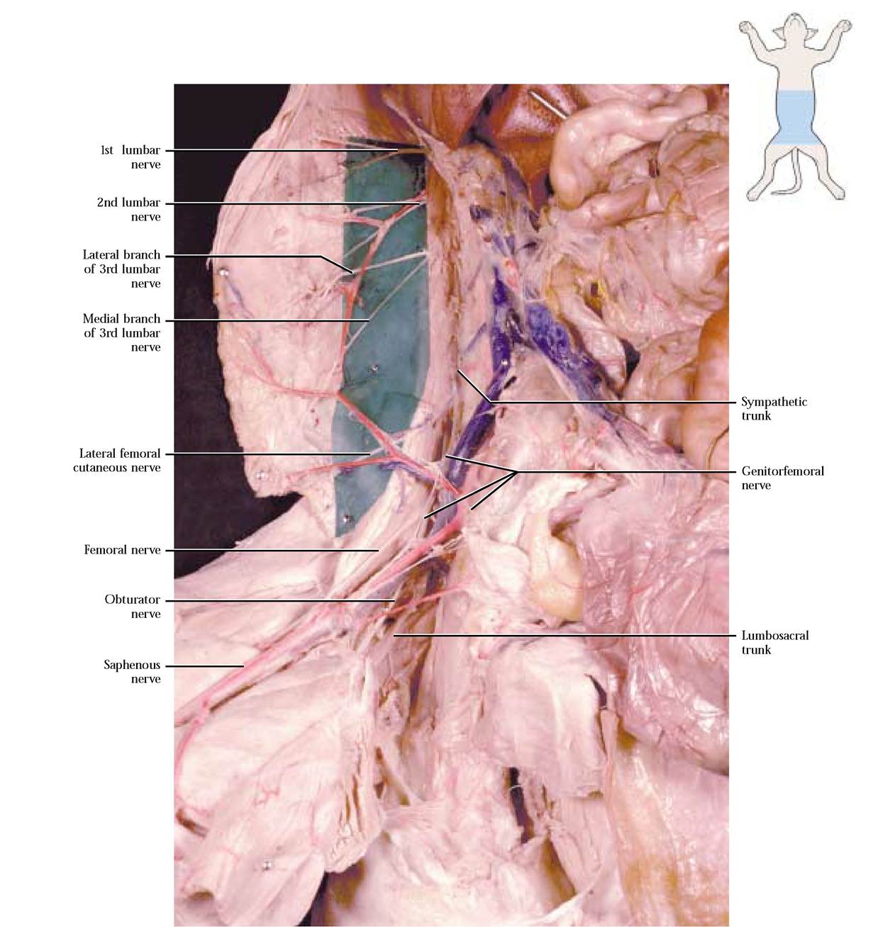
FIGURE 9-10A Lumbosacral plexus: ventral view—enhanced to demonstrate the lumbar nerves.
LUMBOSACRAL PLEXUS (DORSAL VIEW)
The following dissection will expose the nerves of the lumbosacral plexus, nerves originating from the lumbosacral cord, and branches of the sacral nerves and appear in Figure 9-11. During the dissection of the deep hip muscles, many of these nerves were partially exposed.
The largest and most readily observed of these nerves is the ischiatic nerve (sciatic) that extends from the lumbosacral cord. It courses over the lateral muscles of the thigh, giving off a muscular branch that divides to supply the biceps femoris, the semimembranosus, semitendinosus, tenuissimus, and other thigh flexor muscles. As it continues distally, the ischiatic gives off a delicate sural nerve that passes across the gastrocnemius m. to the ankle. As it nears the knee, the ischiatic nerve divides into the common peroneal and tibial nerves [Figure 9-11]. The common peroneal nerve pierces the lateral head of the gastrocnemius m. to supply the peroneal muscles, the tibialis anterior and extensor digitorum
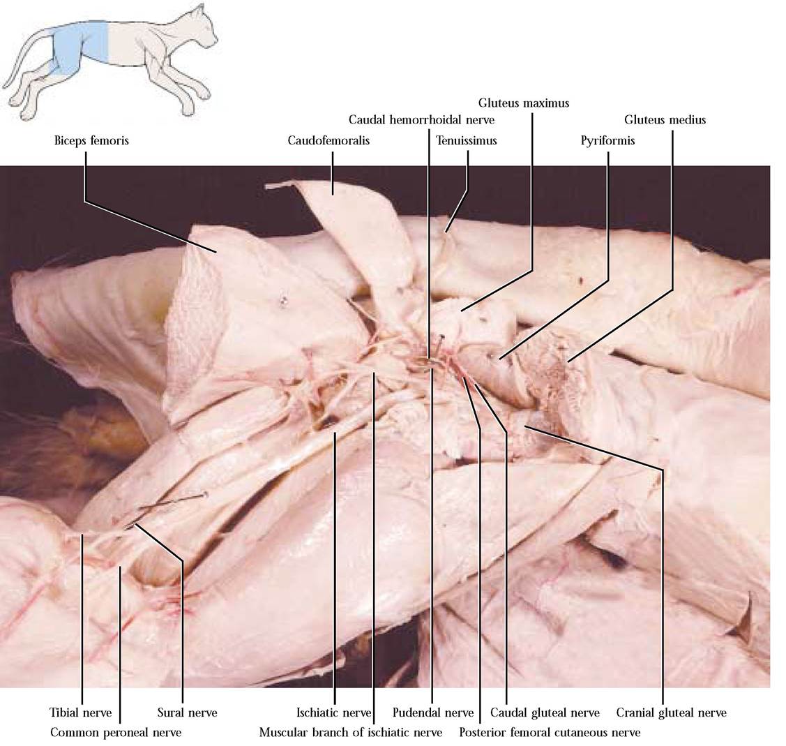
longus muscles, eventually innervating muscles of the digits. The tibial nerve extends between the heads of the gastrocnemius m. and innervates the gastrocnemius, plantaris, soleus, and other muscles associated with extension and flexion of the foot.
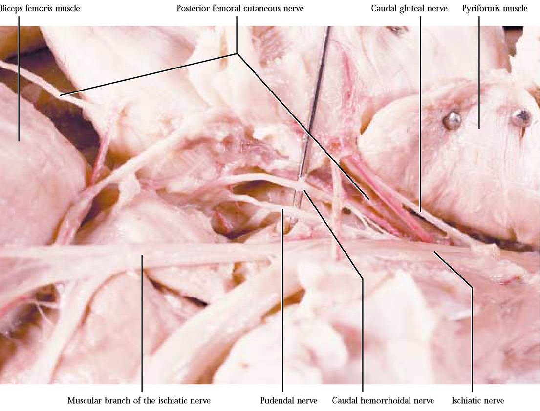
The cranial gluteal nerve, a branch originating from the lumbosacral cord, passes over the dorsal aspect of the ilium and beneath the gluteus minimus m., innervating the gluteus minimus, gluteus medius, gemellus cranialis, and tensor fasciae latae muscles [Figure 9-11]. The caudal gluteal nerve also originating from the lumbosacral cord passes posteriorly
to innervate the caudofemoralis and the gluteus maximus muscles [Figure 9-11 and Figure 9-12]. The posterior femoral cutaneous nerve arises from the second and third sacral nerves and travels with the caudal gluteal blood vessels. It arches over to the biceps femoris muscle and extends over the surface of that muscle innervating the skin at the base of the tail and over the biceps femoris m. [Figure 9-11 and Figure 9-12]. The pudendal nerve arises from the second and third sacral nerves, appearing as a forked nerve that innervates the anus and genital structures in both sexes [Figure 9-11 and Figure 9-12]. The caudal hemorrhoidal nerve, the deepest of these hip nerves, originates from sacral nerves two and three, passes deep, supplying the rectum and the urinary bladder [Figure 9-11 and Figure 9-12]. Generally, this nerve is difficult to find since it lies in connective tissue that must be carefully removed.