Skeletal Muscle Organization
Muscle fibers are arranged in bundles surrounded by fibrous connective tissue. The connective tissue between individual muscle fibers is called endomysium. The sheath surrounding bundles of muscle fibers is called perimysium, and the connective tissue around an entire muscle is known as epimysium.
The epimysium is the deep fascia of the muscle.Muscle fibers may be arranged in parallel sheets, as in the abdominal muscles, or narrower bands, as in the sartorius muscle on the medial side of the thigh; the muscle fibers in these strap muscles are parallel to one another. other arrangements of muscle fibers include fusiform muscles and various pennate, or pen- niform (featherlike), arrangements (Fig. 7-4). in the penniform arrangements, a tendon represents the quill and the muscle fibers attaching to the tendon at an angle represent the vane of the feather. if the fibers come from only one side, the arrangement is called unipennate; from two sides, bipennate; and from three or more sides, multipennate.
The parallel arrangement of muscle fibers in strap muscles provides the greatest potential for overall muscle shortening but is a relatively
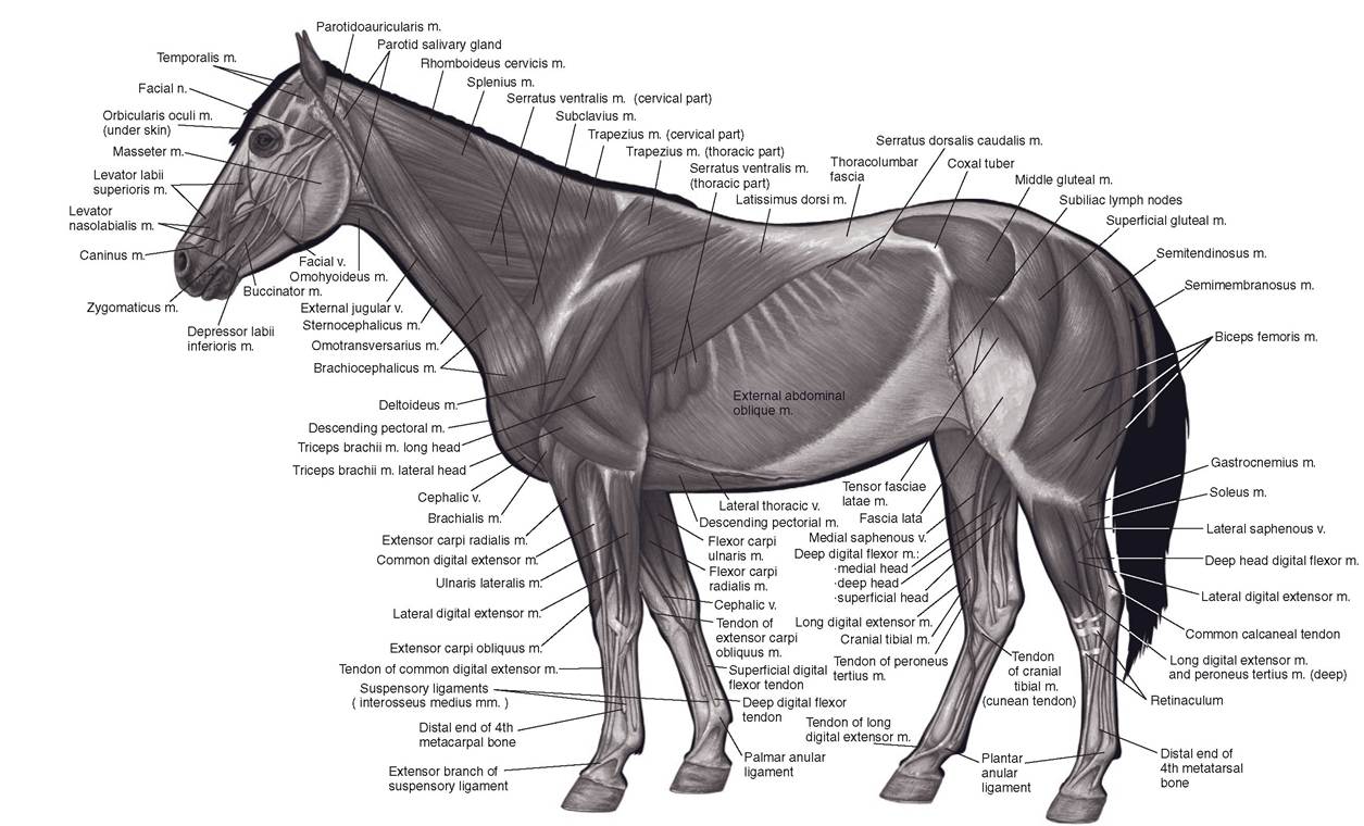
Figure 7-1. Superficial muscles of the horse. (Reprinted with permission of Wiley-Blackwell from McCracken, T.O., Kainer, R.A., and Spurgeon, T.S. Spurgeons Color Atlas of Large Animal Anatomy. Baltimore: Lippincott Williams & Wilkins, 1999.)
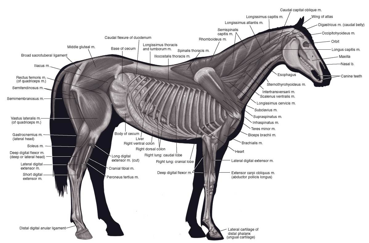
Figure 7-2. Deeper muscles of the horse. (Reprinted with permission of Wiley-Blackwell from McCracken, T.O., Kainer, R.A., and Spurgeon, T.S. Spurgeons Color Atlas of Large Animal Anatomy. Baltimore: Lippincott Williams & Wilkins, 1999.)
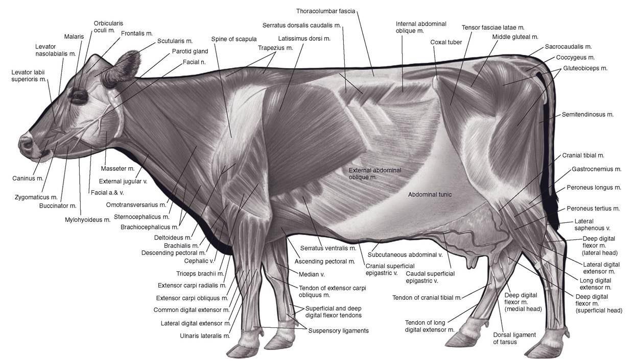
Figure 7-3.
Superficial muscles of the cow. (Reprinted with permission of Wiley-Blackwell from McCracken, T.O., Kainer, R.A., and Spurgeon, T.S. Spurgeon’s Color Atlas of Large Animal Anatomy. Baltimore: Lippincott Williams & Wilkins, 1999.)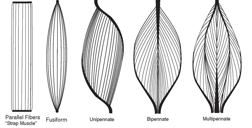
Figure 7-4. Arrangements of muscle fibers: Parallel (a strap muscle), fusiform, unipennate, bipennate multipennate.
weak arrangement, while the pennate arrangement increases the power of a muscle, but at the expense of distance over which it can contract.
Muscle Attachments
if a muscle appears to arise directly from the bone, it is said to have a fleshy attachment. In reality, the muscle fibers attach to very short tendons, which in turn attach to the periosteum of the bone or penetrate the surface of the bone for a short distance.
Tendons, fibrous bands of collagen connecting muscles to bone, are composed of dense, regular connective tissue in parallel bundles. Most tendons are cords or bands that attach spindle-shaped or pennate muscles to bones. Other tendons are flat sheets known as aponeuroses (singular, aponeurosis'), usually associated with flat muscles. The heavy fibrous sheets that cover the muscles of the loin are good examples of aponeuroses.
Most muscles have attachments to two bones (some muscles are attached to soft tissue, e.g., skin). Traditionally, the less mobile attachment is called the origin and the more movable attachment is called the insertion. For example, the m. biceps brachii extends from the scapula to the radius. The scapula usually moves less than the radius during contraction of the biceps, so the origin of the biceps is its attachment to the scapula, and the insertion is its attachment to the radius. in the extremities, the origin usually is proximal and the insertion distal. since the only thing a muscle can actively do is contract, it nearly always tends to bring its origin and insertion closer together, causing one or both of the bones to move.
it is important to note that distinguishing between origin and insertion for some muscles is difficult, as relative motion may change. For example, contraction of the m. brachiocephalicus may advance the thoracic limb when the foot is off the ground, or it may flex the neck to the side if the foot is bearing weight. in such cases, the distinction between origin and insertion is probably a semantic one, without much real anatomical importance.
some muscles have distinctive divisions, called heads, which have separate origins. The triceps is an example of a muscle with multiple heads, in this case three.
Functional Grouping of Muscles
If a muscle is on the side of the limb toward which a joint bends (decreasing the angle between the segments), it is a flexor of that joint. A muscle on the opposite side is an extensor. The m. biceps brachii, on the cranial side of the limb, flexes the elbow. The m. triceps brachii, on the caudal aspect of the limb, takes origin from the scapula and humerus and inserts on the ulna. Thus, the triceps is an extensor of the elbow (Fig. 7-5).
Muscles that tend to pull a limb toward the median plane are adductors, and those that tend to move the limb away from the median plane are abductors. Muscles that pass over more than one joint often have different classifications, depending on the joint on which they are acting. The m. gastrocnemius (the large muscle in the gaskin or calf of the leg) is a flexor of the stifle and an extensor of the hock.
Muscles that encircle an opening, whether they are striated or smooth, are sphincters. The smooth muscle surrounding the opening between the stomach and the intestine forms the pyloric sphincter, which controls passage of food from the stomach. The m. orbicularis oculi is composed of striated muscle fibers in the eyelids, and its contraction squeezes the eyelids shut.
Cutaneous muscles occur in the superficial fascia (a layer of connective tissue) between the skin and the deep fascia covering the skeletal muscles.
These cutaneous muscles attach to the skin and are responsible for movement of the skin. When a fly rests on a horse, the cutaneous muscles enable the horse to shake the skin to dislodge the fly.The muscles involved in a specific action, such as extension of the elbow, may also be classified according to the part each plays in the action. The agonists are the muscles directly responsible for producing the desired action. The antagonists are muscles that oppose that action; they have an action directly opposite that of the agonists. Synergists are muscles that oppose any undesired action of the agonists. For example, in extension of the elbow, a movement produced by contraction of the m. triceps brachii (the agonist for extension of the elbow), the m. biceps brachii and m. brachialis are antagonists because they produce the opposite action, flexion of the elbow. Since the long head of the triceps can flex the shoulder joint as well as extend the elbow, any muscle that opposes flexion of the shoulder joint is a synergist of elbow flexion. The m. supraspinatus and m. brachiocephalicus are synergists for this particular action.
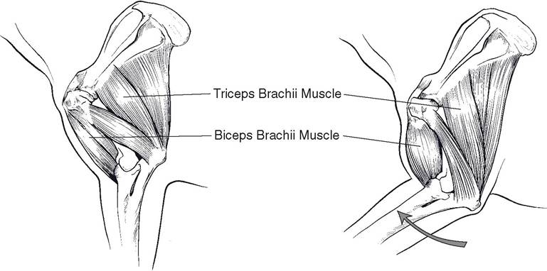
Figure 7-5. Functional grouping of muscles. The m. biceps brachii is a flexor of the elbow, and the m. triceps brachii is an extensor. In this case, the m. biceps brachii is an agonist for elbow flexion, and the m. triceps brachii acts as an antagonist. Any muscles that assist elbow flexion indirectly by stabilizing other joints are considered synergists of that movement.
Whether a given muscle is classified as an agonist, an antagonist, or a synergist depends entirely on the specific action. If flexion (instead of extension) of the elbow is the desired action, the m. biceps brachii and m. brachialis become agonists, and the m. triceps brachii and m. anconeus become antagonists.
Synovial Structures
Synovial structures of the body include joint capsules, bursae, and synovial (tendon) sheaths.
The inner layer of each consists of a connective tissue membrane that produces synovial fluid to reduce friction. Synovial joints were described in detail in Chapter 6.A bursa is a synovial sac between two structures that tend to rub against each other (Fig. 7-6). Clinically important bursae include (1) the bicipital bursa, between the biceps brachii tendon and the proximal end of the humerus; (2) the atlantal bursa, between the ligamentum nuchae and atlas; (3) the supraspinous bursa, between the ligamentum nuchae and the spinous process of the second thoracic vertebra; (4) superficial bursae, between the skin and olecranon process of the ulna at the point of the elbow and between the skin and superficial digital flexor tendon at the point of the hock; and (5) the navicular bursa, between the deep digital flexor tendon and the navicular (distal sesamoid) bone. Normally, a bursa contains only enough fluid to reduce friction between adjacent parts.
Inflammation of a bursa, often associated with excessive fluid, is called bursitis. Enlargement of bursae may be due to trauma, as is usually the case with capped hock, capped elbow (shoe boil), and carpal hygroma. Inflammation followed by formation of a draining tract, which is fairly common, underlies the equine conditions poll evil (at the atlantal bursa) and fi stulous withers (at the supraspinous bursa).
A bursa gives adequate protection to structures that move only a short distance in relation to each other. However, tendons that must travel a long distance (sometimes as much as several inches) over a bone or other structure require protection and friction-free movement for their entire length. This is afforded by a synovial sheath (Fig. 7-6).
A synovial sheath resembles an elongated bursa placed between the tendon and underlying tissue, with the edges of the bursa (sheath) reflected around the tendon until they meet. This results in an inner layer of synovial membrane on the surface of the tendon and a superficial layer of the synovial membrane outside the tendon, forming a closed sac that contains synovial fluid to reduce friction between the tendon and adjacent structures.
The double fold of membrane formed where the edges of the synovial sheath meet is the mesotendon. Vessels and nerves to the tendon reach it by passing through the mesotendon.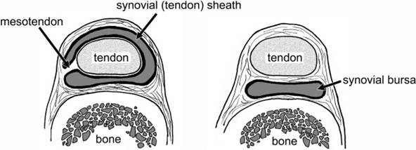
Figure 7-6. Synovial structures. Left) A synovial sheath surrounds a tendon over a greater distance and facilitates its frictionless movement. Reflection of the synovial membrane creates the supporting mesotendon, through which vessels and nerves gain access to the tendon. Right) A bursa lies interposed between a tendon or ligament and a bony prominence.
Inflammation of a synovial sheath and its tendon is called tenosynovitis. Low-grade trauma (for instance, as associated with rigorous schooling of young horses) may result in a mild, painless tenosynovitis of the digital flexor tendon sheath (windpuffs) or the deep digital flexor tendon sheath proximal to the hock (thoroughpin).