Muscles of the Thoracic Limb
See Table 7-1 for list of thoracic limb muscles.
Muscles Acting on the Shoulder Girdle
The scapula and shoulder can make complex movements in humans, but in domestic animals the chief movement of the proximal part of the thoracic limb is a pendulous swing forward and backward.
The muscles that hold the scapula in place contribute to this swinging movement. These muscles are also critical in allowing the weight of the thorax to be supported between the two thoracic limbs (Fig. 77). Superficial muscles of the scapula that link it to the bony thorax include the m. trapezius and the m. omotransversarius. The m. rhomboideus and the m. serratus ventralis are deep.The m. trapezius is a triangular flat muscle that takes origin along the dorsal midline from the head to the lumbar vertebrae. The m. trapezius inserts chiefly on the spine of the scapula. The portion originating cranial to the scapula helps swing the scapula forward; the one attaching behind draws it back.
The m. rhomboideus is a heavier muscle just deep to the trapezius. The m. rhomboideus also originates from the dorsal midline both cranial and caudal to the scapula. The m. rhomboideus inserts on the deep (medial) face of the scapular cartilage.
The m. serratus ventralis is the largest and most important muscle attaching the thoracic limb to the trunk. It is a large, fan-shaped muscle. The origin of the m. serratus ventralis is the widest part and extends from the transverse processes of the cervical and thoracic vertebrae and from the ribs along a curved line just above the sternum as far back as the tenth costal cartilage. The insertion is on the medial side of the dorsal portion of the scapula. The m. serratus ventralis on each side together form a sling that supports the trunk between the thoracic limbs. The cervical portion, on contraction, tends to rotate the distal part of the scapula backward, while the thoracic portion rotates it forward.
The muscle’s cervical attachments allow it to lift the neck as well.The m. omotransversarius is a separate muscle of the shoulder region in most domestic species. It takes origin from the transverse processes of the more cranial cervical vertebrae and inserts on the distal part of the spine of the scapula (clavicular tendon in the horse). With these attachments, the m. omotransversarius usually pulls the distal end of the scapula forward, although with the limb in weightbearing position, it instead assists lateral flexion of the neck.
Muscles Acting on the Shoulder Joint
The shoulder, being a ball-and-socket joint, can make all types of movement. In the quadruped its chief actions are extension and flexion.
Extensors of the Shoulder. The m. brachiocephalicus, as the name implies, extends from the head to the arm. The origin is from the occipital bone of the skull and transverse processes of the cervical vertebrae. It inserts on the lateral side of the proximal part of the humerus proximal to the deltoid tuberosity. The m. brachiocephalicus is the heavy muscle covering the cranial aspect of the point of the shoulder. It
Table 7-1. Muscles of the Thoracic Limb
| Muscle | Origin | Insertion | Primary Actions | Innervation |
| m. trapezius | ligaments of the dorsal midline | scapular spine | lifts, advances, & retracts scapula | accessory n. |
| m. rhomboideus | ligaments of the dorsal midline | medial side scapular cartilage | lifts & advances scapula, lifts neck | cervical & thoracic spinal nn. |
| m. serratus ventralis | transverse processes of caudal cervical & cranial thoracic vertebrae, first 8 or 9 ribs | medial side of scapula & scapular cartilage | supports trunk, lifts neck, advances & retracts scapula | cervical spinal nn, long thoracic n. |
| m. Omotransversarius | bgcolor=white>fascia of shouldertransverse processes of cranial cervical vertebrae | advances limb, lateral neck flexion | cervical spinal nn. | |
| m. brachiocephalicus m. Cleidobrachialis m. cleidocephalicus (m. cleidomastoideus) | occipital bone, transverse processes of cervical vertebrae | medial fascia of arm & forearm, lateral side of humerus, deltoid tuberosity | lateral neck flexion, shoulder extension, advances limb | cervical spinal nn., accessory n. |
| m. latissimus dorsi | spinous processes of thoracic & lumbar vertebrae | medial humerus | limb retraction, shoulder flexion | thoracodorsal n. |
| m. pectoralis superficialis (superficial pectoral m.) | sternum & cranial costal cartilages | medial humerus & fascia of arm & forearm | limb adduction | cranial & caudal pectoral nn. |
| m. pectoralis profundus (deep pectoral m.) | sternum, ribs, abdominal fascia | craniomedial humerus | adduction & retraction of limb, advances trunk when limb is weight-bearing | cranial & caudal pectoral nn. |
| m. subclavius | cranial sternum & costal cartilages | epimysium of m. supraspinatus | support of trunk, shoulder stabilization | cranial pectoral nn. |
| m. supraspinatus | supraspinous fossa of scapula | greater tubercle of humerus | stabilization & extension of shoulder | suprascapular n. |
| m. infraspinatus | infraspinous fossa of scapula | greater tubercle of humerus | stabilization & flexion of shoulder | suprascapular n. |
| m. subscapularis | subscapular fossa | medial humerus | shoulder stabilization | subscapular n. |
| m. teres major | caudal border of scapula | medial humerus (teres major tuberosity) | shoulder flexion | axillary n. |
| m. teres minor | caudal border scapula | proximal to deltoid tuberosity | shoulder flexion | axillary n. |
| m. deltoideus | scapular spine | deltoid tuberosity | shoulder flexion | axillary n. |
| m. coracobrachialis | coracoid process of scapula | medial humerus | shoulder extension | musculocutaneous n. |
| Table 7-1. Continued | ||||
| Muscle | Origin | Insertion | Primary Actions | Innervation |
| m. biceps brachii | supraglenoid tubercle | cranial aspect of proximal radius & ulna | elbow flexion, shoulder extension | musculocutaneous n. |
| m. brachialis | caudal aspect of proximal humerus | medial aspect of proximal radius | elbow flexion | musculocutaneous n. |
| m. triceps brachii | caudal border of scapula, proximal humerus | olecranon process | elbow extension, shoulder flexion (long head only) | radial n. |
| m. anconeus | caudolateral humerus | olecranon process | elbow extension | radial n. |
| m. tensor fasciae antibrachii | caudal border of scapula (via epimysium of triceps m.) | olecranon process, deep fascia of antebrachium | elbow extension, tensing of forearm fascia | radial n. |
| m. extensor carpi radialis | lateral humeral epicondyle | cranial aspect of proximal metacarpus | carpal extension | radial n. |
| m. extensor carpi obliquus | craniolateral radius | medial metacarpus | carpal extension | radial n. |
| m. extensor digitorum communis (common digital extensor m.) | lateral humeral epicondyle | distal phalanx | digital extension, carpal extension | radial n. |
| m. extensor digitorum lateralis (lateral digital extensor m.) | lateral collateral ligament of elbow, proximal radius & ulna | dorsal aspect of digit | digital extension, carpal extension | radial n. |
| m. extensor carpi ulnaris (m. ulnaris lateralis) | lateral humeral epicondyle | lateral metacarpus, accessory carpal bone | carpal flexion (?) | radial n. |
| m. pronator teres | medial humeral epicondyle | medial aspect proximal radius | elbow flexion | median n. |
| m. flexor carpi radialis | medial humeral epicondyle | palmeromedial aspect of proximal metacarpus | carpal flexion | median n. |
| m. flexor carpi ulnaris | medial humeral epicondyle, olecranon | accessory carpal bone | carpal flexion | ulnar n. |
| m. flexor digitorum superficialis (superficial digital flexor m.) | medial humeral epicondyle | middle phalanx | digital flexion, carpal flexion | ulnar n. |
| m. flexor digitorum profundus (deep digital flexor m.) | medial humeral epicondyle, caudal radius, medial olecranon | palmar surface of distal phalanx | digital flexion, carpal flexion | median & ulnar nn. |
n(n), nerve(s)
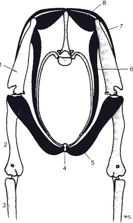
Figure 7-7. Muscular suspension of the thorax between the thoracic limbs. 1, Scapula; 2, humerus; 3, radius and ulna; 4, sternum; 5, m. pectoralis profundus; 6, m. serratus ventralis; 7, m. trapezius; 8, m. rhomboideus. The attachment of limbs to the trunk is achieved through a synsarcosis, rather than by a bony joint.
raises and advances the shoulder. The m. brachiocephalicus is also the principal extensor of the shoulder and acts as a lateral flexor of the neck when the limb is weight bearing.
The m. brachiocephalicus is subdivided into the m. cleidobrachialis, extending from the clavicular tendon (representing the vestigial clavicle) to the humerus, and the m. cleidocephalicus, extending from the clavicular tendon to the head and neck. in species other than the horse, the m. cleidocephalicus may be further subdivided into a mastoid part attaching to the mastoid process of the temporal bone and either an occipital part (in ruminants and pigs) or a cervical part (in carnivores). in horses, the sole attachment of the m. cleidocephalicus is to the mastoid process, and in this species the muscle is often called the m. cleidomastoideus. The m. cleidobrachialis is comparable to the anterior deltoid muscle of humans.
The m. supraspinatus originates from the supraspinous fossa of the scapula cranial to the spine. it inserts on the greater tubercle (both greater and lesser in the horse) of the humerus. The m. supraspinatus may assist in extending the shoulder but acts chiefly as a stabilizing ligament of the shoulder joint. This is one of the muscles that atrophies (shrinks) in sweeny in horses, a condition that results from damage to its motor innervation, the suprascapular nerve.
Flexors of the Shoulder. The m. teres major originates from the dorsal part of the caudal border of the scapula and inserts on the teres major tuberosity on the medial side of the shaft of the humerus. it is a strong flexor of the shoulder joint.
The m. latissimus dorsi is a wide, triangular muscle that originates from the spinous processes of the thoracic and lumbar vertebrae by means of a wide aponeurosis, the thoracolumbar fascia. It inserts with the m. teres major on the medial side of the humerus and is a strong flexor of the shoulder. Also, it pulls the thoracic limb caudad or, if the limb is fixed, advances the trunk.
The m. infraspinatus originates from the infraspinous fossa just caudal and ventral to the spine of the scapula. it inserts into the caudal part of the greater tubercle of the humerus. The m. infraspinatus also acts as a strong collateral ligament of the shoulder joint and may abduct, flex, and outwardly rotate the shoulder. This muscle also atrophies in cases of sweeny.
The m. teres minor lies just distal to the infraspinatus muscle and has the same action as the m. infraspinatus. The m. teres minor originates from the distal caudal border of the scapula and inserts on the teres minor tuberosity of the humerus just distal to the greater tubercle of the humerus.
The m. deltoideus extends from the spine of the scapula to the deltoid tuberosity of the humerus. it is an abductor and flexor of the shoulder joint.
Adductors of the Shoulder. The pectoral muscles form the substance of the brisket. They originate from the sternum and insert mainly on the proximal part of the humerus. Commonly, they are divided into the superficial pectoral muscle (m. pectoralis superficialis) and the deep pectoral muscle (m. pectoralis profundus). These pectoral muscles are strong adductors of the forelimb, and the deep pectoral muscle also advances the trunk when the limb is fixed on the ground (weight bearing).
The m. subclavius is absent in carnivores, small in ruminants, and well developed in horses and pigs. It has sometimes been considered part of the deep pectoral muscle. The m. subclavius arises from the cranial sternum and costal cartilages and arcs craniodorsad to insert on the epimysium of the m. supraspinatus. This muscle is part of the support of the trunk and contributes to stabilization of the shoulder joint.
The m. coracobrachialis is a small muscle extending from the coracoid process on the medial side of the scapula to the medial side of the shaft of the humerus. The location of the muscle belly suggests a shoulder flexor, but its attachments make this muscle an extensor of that joint.
The m. subscapularis stabilizes the shoulder on the medial side. It originates from the subscapular fossa on the medial side of the scapula below the attachments of the m. rhomboideus and m. serratus ventralis. It inserts on the lesser tuberosity of the humerus and provides some adduction to the shoulder joint.
Muscles Acting on the Elbow
Since the elbow is a hinge joint, the muscles acting on it are either flexors or extensors. In quadrupeds, the extensors are stronger than the flexors because they support the weight of the body by maintaining the limbs in extension (Fig. 7-8).
Extensors of the Elbow. The m. triceps brachii has three heads. The long head originates from the caudal border of the scapula, and the medial and lateral heads originate from the respective sides of the humeral diaphysis. carnivores have an accessory head that also originates from the humerus between the medial and lateral heads (although this arrangement gives the carnivore four heads on this muscle, it is still called a triceps). All heads insert on the olecranon process of the ulna. The triceps is the strongest extensor of the elbow. The long head may also act to flex the shoulder.
The m. anconeus, deep to the m. triceps brachii, is a rather small muscle that covers the caudal aspect of the joint capsule of the elbow. It also originates on the humerus, inserts on the olecranon process, and extends the elbow.
The m. tensor fasciae antibrachii originates via a thin aponeurosis that is blended with the long head of the triceps muscle and the m. latissimus dorsi. The flattened muscle belly lies on the caudomedial aspect of the arm and inserts via a second aponeurosis on the olecranon and antebrachial fascia. The muscle’s name reflects its action on the antebrachial fascia (it tenses it), but through these fascial connections the m. tensor fasciae antibrachii also assists the triceps in extension of the elbow.
Flexors of the Elbow. The m. biceps brachii originates on the supraglenoid tubercle just dorsal and cranial to the articular surface of the scapula. It inserts on (1) the radial tuberosity on the cranial aspect of the proximal radius, (2) the medial collateral ligament of the elbow, and (3) the antebrachial fascia. The tendinous blending with the antebrachial fascia forms a palpable cordlike structure on the flexor surface of the elbow called the lacertus fi brosus. The biceps assists in holding the shoulder joint in apposition and may extend it to some extent. However, the chief action of the m. biceps brachii is flexion of the elbow.
The m. brachialis is strictly a flexor of the elbow since it originates on the humerus and inserts on the cranial aspect of the radius (and in some species the ulna).
As ungulates have only limited or absent ability to pronate and supinate their limbs, the m. pronator teres is reduced to a ligament in horses and only a small, weak muscle in ruminants and pigs. In these species, it acts primarily as a flexor of the elbow. It originates on the medial epicondyle of the humerus and inserts on the medial side of the radius.
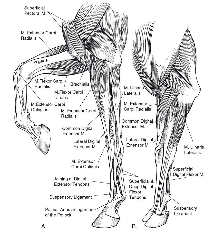
Figure 7-8. Muscles of the thoracic limb. A) Equine. B) Bovine.
Extensor muscles of the carpus and digit (discussed in the next section), which originate on the lateral epicondyle of the humerus, may assist in flexion of the elbow as a secondary function.
Muscles Acting on the Carpus
The carpus, like the elbow, acts essentially as a hinge joint. The extensors of the carpus lie on the craniolateral aspect of the limb, and the flexors are found on the caudomedial side.
Extensors of the Carpus. The m. extensor carpi radialis is the largest extensor of the carpus. It extends from the lateral epicondyle of the humerus to the proximal end of the metacarpal region. It inserts on the metacarpal tuberosity on the dorsal surface of the proximal end of the metacarpus. This is the most prominent muscle on the front of the forearm and is the most cranial muscle of the group. As the name implies, the m. extensor carpi radialis acts primarily as an extensor of the carpus.
The m. extensor carpi obliquus is a flat, triangular extensor of the carpus lying deep to the digital extensor muscles of the antebrachium. It arises from the craniolateral aspect of the distal half of the radius (and ulna in species with a complete ulna). Its oblique tendon crosses mediad on the cranial aspect of the carpus, superficial to the tendon of the m. extensor carpi radialis, to insert on the most medial metacarpal bone, which is the second in the horse, the third in the cow and sheep, and the second in pigs. In humans, this muscle is one of the well-developed abductors and extensors of the thumb (the pollex) and so in this species is called the m. abductor pollicis longus. This name is only infrequently used in veterinary anatomy, since most domestic species lack a well-developed first digit.
The m. extensor carpi ulnaris (formerly m. ulnaris lateralis) is the most caudal of the extensor muscles. It also takes origin from the lateral epicondyle of the humerus, but passes downward over the lateral side of the carpus to insert on the most lateral metacarpal bone. In most domestic animals, this muscle probably produces weak flexion of the carpus, although by origin and nerve supply it belongs with the extensor group. It also produces some outward rotation of the forearm.
In addition to these carpal extensors, the extensor muscles of the digits whose tendons pass over the dorsal surface of the carpus may act secondarily as extensors of the carpus.
Flexors of the Carpus. On the medial side of the forearm, the m. flexor carpi radialis is just caudal to the radius, which is palpable directly beneath the skin. It takes origin from the medial epicondyle of the humerus and inserts on the palmar aspect of the proximal end of the metacarpus (medial side).
on the caudal side, the m. fl exor carpi ulnaris exerts considerable leverage as a flexor of the carpus by inserting on the accessory carpal bone, which projects in a palmar direction from the lateral aspect of the carpus.
These muscles are, of course, primarily flexors of the carpus, but they may act slightly in extension of the elbow. Recall, too, that despite its common name, the m. extensor carpi ulnaris appears to be a flexor of the carpus in domestic animals (for this reason, many veterinary anatomists recommend the alternate name, m. ulnaris lateralis).
Muscles Acting on the Digits
Extensors of the Digit. The common digital extensor muscle (m. extensor digitorum communis) is the longest extensor muscle in the thoracic limb. It originates from the lateral epicondyle of the humerus close to the m. extensor carpi radialis. Its tendinous insertion is on the extensor process of the distal phalanx and on the proximal ends of the middle and proximal phalanges. The tendon is single in the horse; double in the cow, sheep, and goat; and split into four separate tendons in the pig and carnivores, in which species it inserts on the second through the fifth digits. This muscle is an extensor of all joints of the digit, including the fetlock. It may also assist in extending the carpus and even in flexing the elbow (owing to its origin on the humerus).
The common digital extensor m. of animals with more than a single digit has several distinct heads. In ruminants, one of these heads gives rise to its own tendon that inserts on the third digit (the medial toe). This head of the common digital extensor m. is sometimes identified as a separate muscle, the medial digital extensor muscle.
The lateral digital extensor m. (m. extensor digitorum lateralis) is found in all species. Its origin is just caudal to the common digital extensor m. on the lateral collateral ligament of the elbow and lateral radius and ulna. The insertion varies according to the number of digits present, and there are interspecific differences regarding to which phalanx the tendon attaches. in pigs it inserts on the fourth and fifth digits; in ruminants, on the middle phalanx of the fourth digit; and in the horse, on the proximal phalanx of the third (and only) digit.
Flexors of the Digit. In all animals the principal digital flexors are the superficial and the deep digital flexor muscles. The deep digital flexor m. (m. flexor digitorum profundus) lies the closest to the metacarpal bones. it originates from the humerus, radius, and ulna. its long, stout tendon extends distad through the carpal canal, then along the palmar side of the metacarpus to insert on the palmar surface of the distal phalanges. As with the common digital extensor tendon, the number of insertions depends on the number of digits, with the main tendon dividing into individual slips, one per digit, just proximal to the fetlock. The deep digital flexor is the only muscle that flexes the distal interphalangeal joint. secondarily, it also flexes the more proximal joints of the digit and the carpus. The deep digital flexor m. also is important in supporting the fetlock.
The superficial digital flexor m. (m. flexor digitorum superficialis) is similar to the deep digital flexor m., but it inserts primarily on the proximal part of the middle phalanx of each digit. in the horse, the superficial digital flexor tendon inserts on the palmar aspects of the proximal end of the middle phalanx and the distal end of the proximal phalanx. Tendons of both the superficial and deep digital flexor mm. can be palpated palmar to the cannon bone. The expression bowed tendons describes a traumatic condition of horses involving tendonitis of one or both of these tendons in the cannon region (Fig. 7-9).
Interosseous muscles lie between the metacarpal bones of carnivores and humans. in large ungulates most of the muscle tissue has been
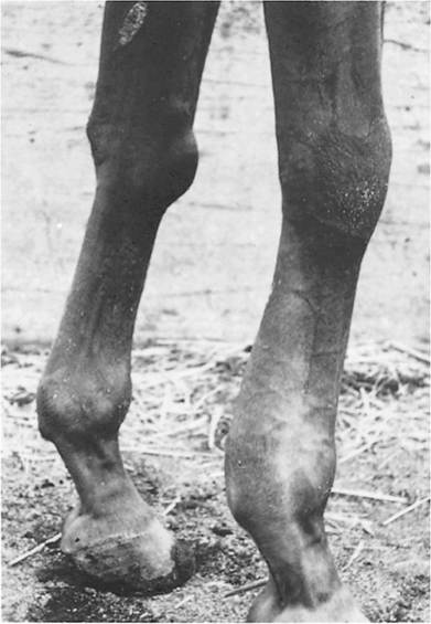
Figure 7-9. Tenosynovitis (bowed tendon). Note swelling on the palmar aspect above the fetlock in the area of the flexor tendons.
replaced with connective tissue, and these structures are known as the suspensory ligaments. They take origin from the palmar aspect of the proximal metacarpus and insert on the proximal sesamoid bones. The suspensory ligaments are part of the passive support of the metacarpophalangeal joint.