SKELETAL SYSTEM
Amphibians
There is significant diversity of skeletal elements among amphibians. Caecilians lack pectoral and pelvic girdles, as well as the sacrum. Locomotion in this group is primarily achieved through worm-like regional contraction of the body (vermiform motion), or lateral, eel-like undulations (Stebbins & Cohen 1995; Wright 2001c).
Salamanders (Fig. 1.9) typically have four limbs, though the hindlimbs are greatly reduced in the mud eels (Amphiuma spp.) and missing in sirens (Siren spp. and Pseudobranchus spp.) (Stebbins & Cohen 1995; Wright 2001c). Generally, four toes are present on the forefoot and five on the hind, although this is variable between species. Salamanders are capable of regenerating lost toes and limbs. Cleavage planes, or predetermined zones of breakage, are present in the tails of many species so that when the animal is threatened or injured the tail breaks free of the body. This is known as autotomy; the lost tail will regenerate (Stebbins & Cohen 1995).
Anurans have several adaptations for saltatory locomotion or jumping. They have four limbs, and the hind legs are elongated (Fig. 1.10). There are generally four toes on the forefoot and five on the hind foot. The vertebrae are fused and the vertebral column is divided into the presacral, sacral, and postsacral regions. The sacrum itself is not present, and the pelvic girdle is fused. The forelimb is composed of the humerus, a fused radio-ulna, carpals, metacarpals, and phalanges, and the hind limb is formed by the femur, fused tibiofibula, tarsals, metatarsals, and phalanges. Caudal vertebrae are replaced by a fused
Amphibians
8
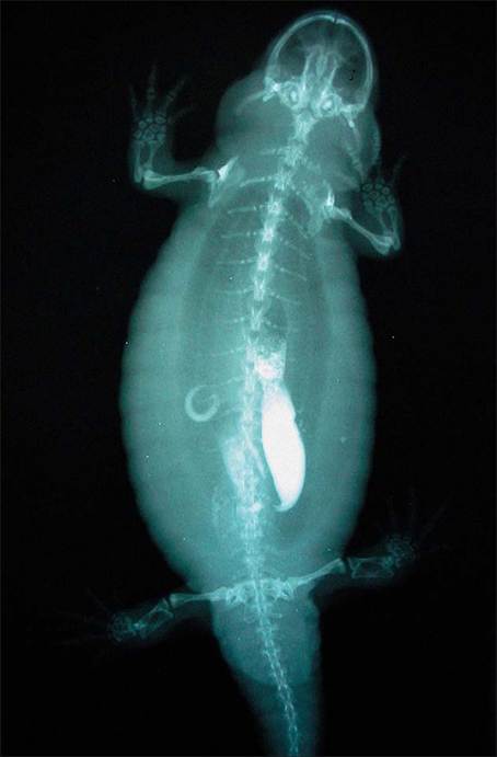
Figure 1.9 • Dorsoventral projection of gastrointestinal contrast study of a salamander. The radiograph is normal.
(Photo by Whiteside.)urostyle. Tadpoles can regenerate limbs, but adult anurans generally cannot (Wright 2001c).
CARDIOVASCULAR SYSTEM
The amphibian cardiovascular system is comprised of the arterial, venous, and well-developed lymphatic structures. The amphibian heart is 3-chambered, with two atria and one ventricle. The interatrial septum is fenestrated in caecilians and most salamanders, but complete in anurans, allowing varying degrees of mixture of oxygenated and deoxygenated blood (Wallace et al. 1991; Wright 2001c).
Blood draining from the caudal half of amphibians passes through the kidneys prior to entering the postcaval vein.
CLINICAL NOTE
Recent studies in reptiles have demonstrated little effect of the renal portal system on pharmacokinetics of drugs administered in the caudal half of the body (Holz et al. 1999, 2002); however, until similar studies are performed on amphibians it is advisable to avoid administration of medications in the hind limb or tail (if present) of amphibians.
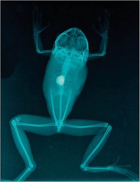
Figure 1.10 • Dorsoventral projection of a Red-eyed tree frog (Agalychnis callidryas). Note the fracture of the right femur, as well as the radio-opaque gastric foreign body. (Photo by Helmer.)
Amphibian lymph consists of all the components of blood, with the exception of erythrocytes. The lymphatic system includes lymph hearts (also known as lymph sacs or lymph vesicles) that beat independently of the heart at a rate of 50-60 beats per minute. These structures ensure unidirectional flow of lymph back to the heart (Wright 2001c).
Venepuncture sites
The choice of venepuncture sites will depend on the size and species of the patient. In anurans, potential sites include the:
heart (cardiocentesis)
ventral abdominal vein (often visible percutaneously in larger frogs) (Fig. 1.11)
■ femoral vein
■ lingual vein
In salamanders the ventral tail vein is readily accessible (Whitaker & Wright 2001).