Skin
Skin covers the outside of the animal and is continuous with mucous membranes at oral, anal, and urogenital orifices, the vestibule of the
nostril, and the palpebral fissure; these sites are characterized by a mucocutaneous junction.
Thickness of skin varies both between species and on a given individual, being generally thickest where it is most exposed (e.g., on the back) and thinner in protected regions (e.g., the groin). The skin adheres tightly to underlying structures in some locations, but in others is loosely attached to allow for considerable movement. The looseness of skin attachment is exploited by veterinarians who frequently inject medications or fluids for rehydration into the space underneath the skin (a subcutaneous injection).Epidermis
The outer layer of skin, the epidermis, is an avascular stratified squamous epithelium that is nearly free of nerve endings. in most areas it can be divided into several histologic layers (Fig. 131). They are as follows, from deep to superficial: (1) A layer of mitotically active cuboidal or columnar cells, the stratum basale, follows the contour of the underlying dermis, to which it is closely applied. (2) The stratum spinosum has a spiny appearance because of its desmosomes (intercellular bridges) connecting adjacent cells. (3) The stratum granulosum consists of spindleshaped cells containing basophilic keratohyalin granules. (4) The stratum lucidum, which is variably present, comprises cells that are poorly stainable. (5) The stratum corneum consists of layers of dead, flattened cells. Presence and relative thickness of each layer is reflected in the overall thickness of the skin (Fig. 13-1).
Cells in the stratum basale undergo mitotic division, which pushes the more superficial layers still farther from the blood vessels in the underlying dermis. As distance from nutrients increases, the cells flatten and die, leaving a dense mat of their primary constituent, the fibrous protein keratin.
The drying and hardening of the superficial cells, a process called both keratinization and cornification, renders the surface of the skin tough and resistant to drying. As the stratum basale continuously adds cells to overlying layers, the stratum corneum flakes off and is replaced. The rate at which this occurs can be influenced by trauma or disease processes. A callus is a local increase in thickness in response to continuous trauma.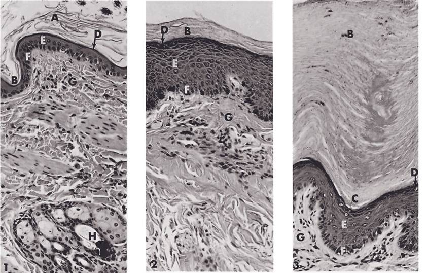
Figure 13-1. Feline skin. 1) Hairy skin from lumbar region. Note multiple hair follicles near bottom of micrograph. 2) Hairless skin from nose; epidermis is thicker in this region. 3) Footpad. Epidermis is markedly thickened in this region, with most of the increase due to thickening of the stratum corneum. A, Flaking keratin; B, stratum corneum; C, stratum lucidum; D, stratum granulosum; E, stratum spinosum; F, stratum basale; G, dermis; H, hair follicle. (Reprinted with permission of Wiley-Blackwell from Dellmann H.D. and Brown E.M. Textbook of Veterinary Histology. 3rd ed. Philadelphia: Lea & Febiger, 1981.)
Dermis
The epidermis forms an undulating sheet with fingerlike projections, the epidermal pegs, which project into the underlying connective tissue, the dermis. The dermis (also known as the corium, especially where it is associated with modifications like hoofs and horns) bears ridges and nipplelike projections (the dermal papillae) that interdigitate with the overlying epidermis (Fig. 13-2); these are most especially prominent in weight-bearing structures, such as footpads and hoofs. The interface between epidermal pegs and dermal papillae provides increased surface area for formation of a strong junction between these two layers. A blister is a local disruption of this association between layers, usually due to repeated trauma or thermal injury.
Arteries, veins, capillaries, and lymphatics of the skin are contained in the dermis.
Sensory nerve fibers, in addition to supplying the dermis, may extend a short distance into the epidermis. Sympathetic nerves provide motor innervation to blood vessels, glands, and arrector pili muscles of hair follicles in the dermis. These structures do not receive parasympathetic innervation.Color of skin is due to the pigment granules generated in the cytoplasm of the resident pigment cells, melanocytes. These cells in the stratum basale produce the pigment, melanin, which is brown, yellowish-brown, or black. Packets of melanin pigment are manufactured by the melanocytes and transferred to surrounding cells of the epidermis; the same process incorporates pigment into cells that cornify into hairs. The expression of different colors in skin and hair comes primarily from the relative amount of melanin produced in the melanocytes rather than from differences in numbers of melanocytes or presence of other pigments. This expression can be influenced by certain pituitary hormones, notably melanocyte-stimulating hormone (MsH) and adrenocorticotropic hormone (ACTH) (see Chapter 12).
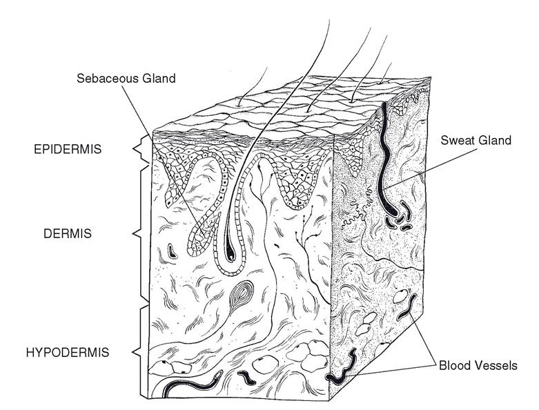
Figure 13-2. Skin anatomy.
Absence of pigment in the skin (albinism), which may be partial or total, arises from a genetic inability of melanocytes to manufacture pigment. Lack of pigment can render the skin and surface mucous membranes more susceptible to actinic damage (i.e., cellular damage due to ultraviolet light), hence formation of carcinoma (cancer) of the skin or other exposed epithelia. Cancer eye, or squamous cell carcinoma of the conjunctiva of the eye, is common in white-faced cattle (e.g., Hereford) living at high elevations, where the ultraviolet component of sunlight is little attenuated.
Hypodermis
in nearly all areas of the body, a layer of loose connective tissue separates the dermis from underlying structures. This areolar connective tissue, known variously as the superficial fascia, subcutis, or hypodermis, permits movement of the skin without tearing. Where the skin is tightly attached to underlying bone or muscle, a dimple on the body surface may be seen. This is a “tie,” as is seen where the dermis is attached to the spinous processes of vertebrae. Variable amounts of fat, the panniculus adiposus, are present in the hypodermis, with species-dependent distribution and relative abundance. The panniculus adiposus is an especially notable feature of pigs; on the dorsum of the pig, it is called backfat.