Small Intestine
The duodenum is the first of three divisions of the small intestine (Figs. 20-12 to 20-14). It is closely attached to the right side of the dorsal body wall by a short mesentery, the mesoduodenum.
The duodenum arises at the pylorus of the stomach and receives ducts from the pancreas and liver in this region. It passes caudad on the right side of the abdominal cavity toward the pelvic inlet, then crosses to the left side caudal to the root of the great mesentery (discussed later) and reflects craniad to join the jejunum. The mesentery becomes considerably longer at this duodenojejunal flexure, and the duodenum is attached at this site to the descending colon by a serosal ligament, the duodenocolic fold.The transition between duodenum and the next portion of the small intestine, the jejunum, is defined by the marked increase in the length of the supporting mesentery. The jejunum is the longest part of the small intestine (e.g., as much as 28 m in the horse). Histologically, the jejunum is similar to the duodenum, although lymph nodules at the mucosal-submucosal junction may be more numerous.
The ileum is the short last part of the small intestine. It is distinguished from the jejunum by a fold of mesentery between it and the cecum. This ileocecal fold is found on the side of the intestine opposite the attachment of the mesentery (the antimesenteric side). The lumen of the ileum communicates with that of the large intestine at the ileal orifice; this junction is found in the right caudal part of the abdominal cavity in all species. The ileal epithelium features numerous goblet (mucous) cells, and aggregates of lymph nodules in this region are more abundant than in other parts of the small intestine. Their especially prominent arrangement in the ileum has led to the use of the term Peyer’s patches to distinguish them.
The mesenteries that suspend the small intestine from the dorsal body wall can be named according to the part of the intestine supported, that is, mesoduodenum, mesojejunum, and mesoileum.
The mesoduodenum is generally short, so the location of the duodenum is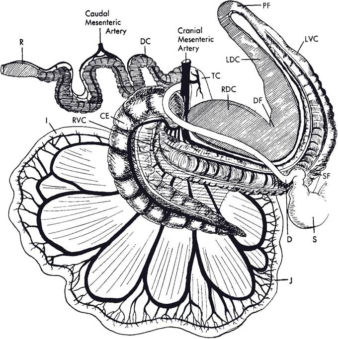
Figure 20-12. Gastrointestinal tract of the horse. S, Stomach; D, duodenum; J, jejunum; I, ileum; CE, cecum; RVC, right ventral colon; SF, sternal flexures; LVC, left ventral colon; PF, pelvic flexure; LDC, left dorsal colon; DF, diaphragmatic flexure; RDC, right dorsal colon; TC, transverse colon; DC, descending colon; R, rectum. (Redrawn after Nickel R., Schummer A., and Seiferle E. Lehrbuch der Anatomie der Haustiere. Berlin: Paul Parey, 1960. Permissions from Wiley-Blackwell.)
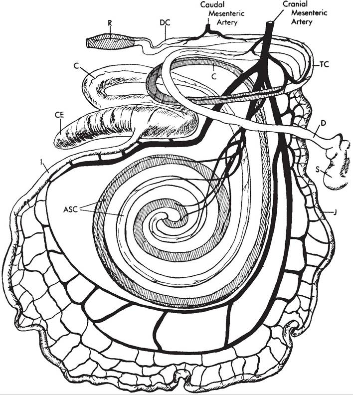
Figure 20-13. Gastrointestinal tract of the ox. S, Stomach (abomasum; forestomach not shown); D, duodenum; J, jejunum; I, ileum; CE, cecum; C, proximal loop and distal loop; ASC, ansa spiralis; TC, transverse colon; DC, descending colon; R, rectum. (Redrawn after Nickel R., Schummer A., and Seiferle E. Lehrbuch der Anatomie der Haustiere. Berlin: Paul Parey, 1960. Permissions from Wiley-Blackwell.)
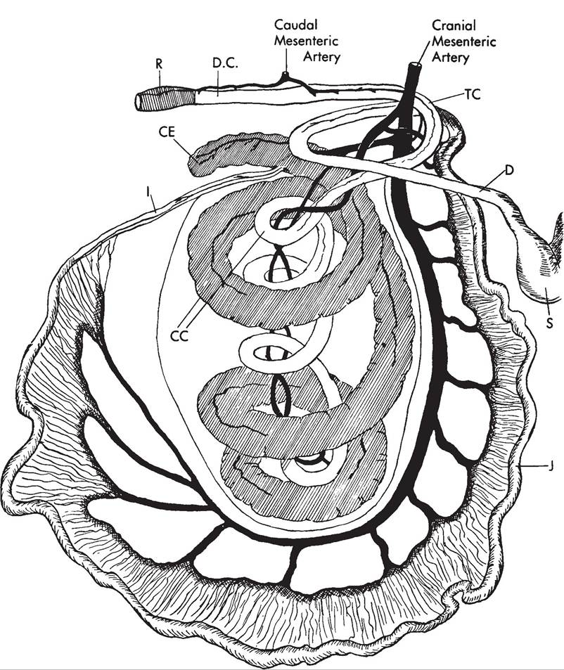
Figure 20-14. Gastrointestinal tract of the pig. S, Stomach; D, duodenum; J, jejunum; I, ileum; CE, cecum; CC, ansa spiralis (coiled colon); TC, transverse colon; DC, descending colon; R, rectum. (Redrawn after Nickel R., Schummer A., and Seiferle E. Lehrbuch der Anatomie der Haustiere. Berlin: Paul Parey, 1960. Permissions from Wiley-Blackwell.)
relatively fixed. The mesenteries supporting the jejunum and ileum, on the other hand, are long and fanlike, so that many meters of intestine are connected to a small region of the dorsal body wall. The mesojejunum and mesoileum are often collectively called the great mesentery, and the narrow stalk by which they are attached to the body wall and through which blood vessels, nerves, and lymphatics reach the intestines is commonly called the root of the great mesentery. The length of the great mesentery permits considerable mobility of the intestinal mass.