Ruminant Stomach
The ruminant stomach is actually a single stomach modified by marked expansion of the esophageal region into three distinct and voluminous diverticula, the rumen, reticulum, and omasum, collectively known as the forestomach.
These are lined with nonglandular stratified squamous epithelium and comprise a series of chambers where food is subjected to digestion by microorganisms before passing through the digestive tract to the smaller glandular portion of the stomach in the ruminant, the abomasum. The stomach of the ox is shown in Figures 20-9, 20-10, and 20-11.Ruminoreticulum
Because of their functional and anatomic relatedness, the reticulum and rumen are often collectively called the ruminoreticulum. The opening of the esophagus (the cardia) is about the level of the middle of the seventh intercostal space, and it opens into the dorsal space that is common to both the rumen and reticulum. The mucosa in the region of the cardia forms two heavy muscular folds that together create a groove extending from the cardia to omasum. This is the sulcus ruminoreticularis (variously called the esophageal, gastric, or reticular groove). in nursing ruminants, the act of suckling initiates a reflex contraction of the muscular walls of the sulcus, transforming it from a groove to a closed tube that connects the cardia with the omasum. By this reflex, swallowed milk bypasses the ruminoreticulum and is instead delivered to the more distal parts of the stomach; this ensures that the milk will not be allowed to sour in the forestomach.
The reticulum is the most cranial compartment of the forestomach. its mucosa is thrown up into intersecting ridges that give the reticulum its common name, the “honeycomb.” Foreign objects such as wire or nails that are
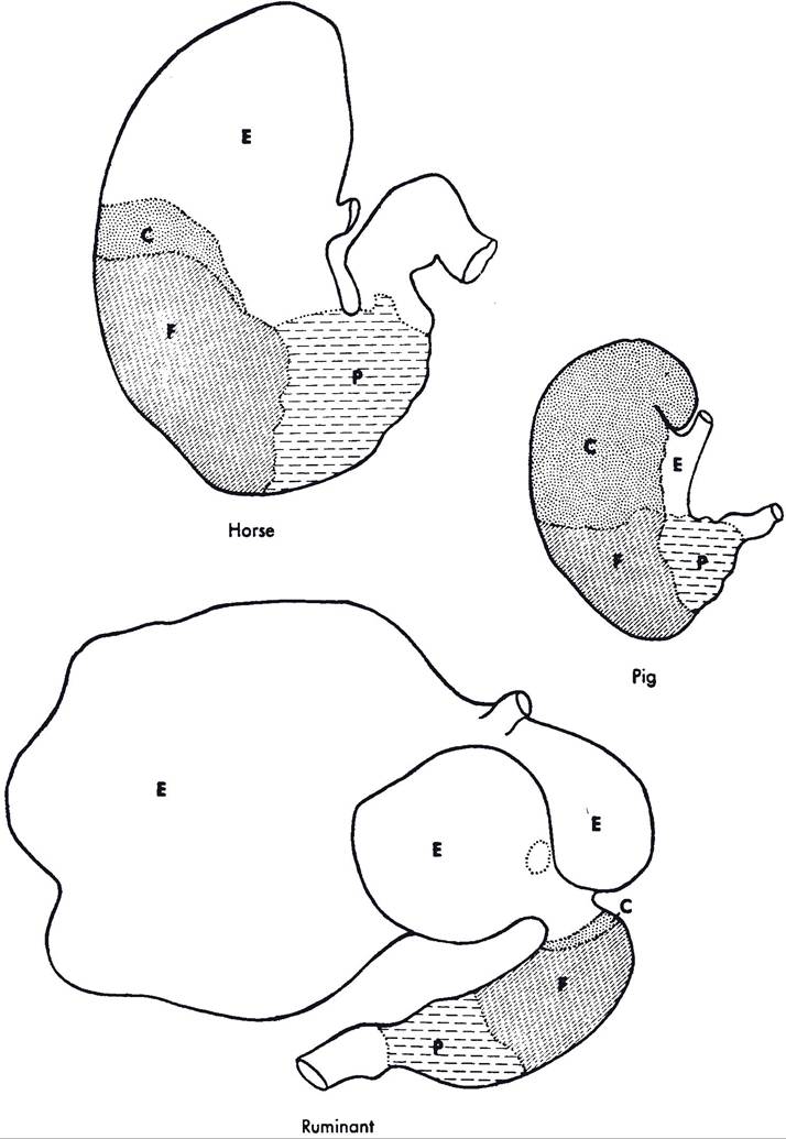
Figure 20-9.
Stomach regions as defined by glandular type in the horse, pig, and ruminant. E, esophageal region; C, cardiac gland region; F, fundic gland region; P, pyloric gland region.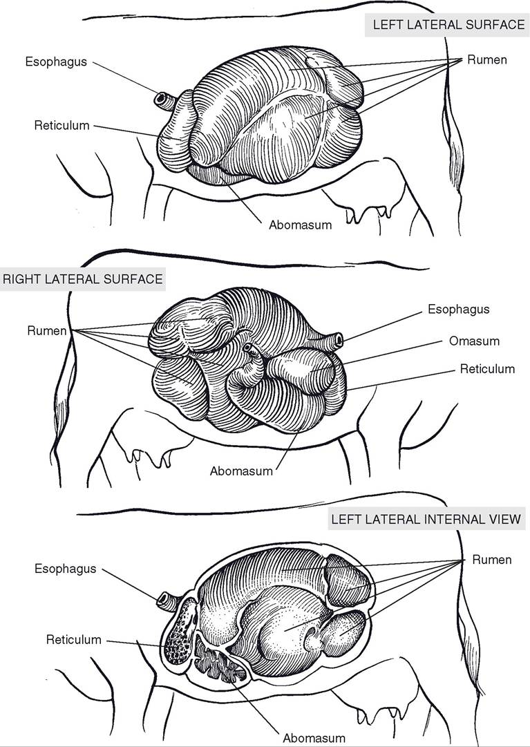
Figure 20-10. Bovine stomach in situ. Top) View from left. Center) View from right. Bottom) Internal anatomy of the ruminoreticulum as viewed from the left.
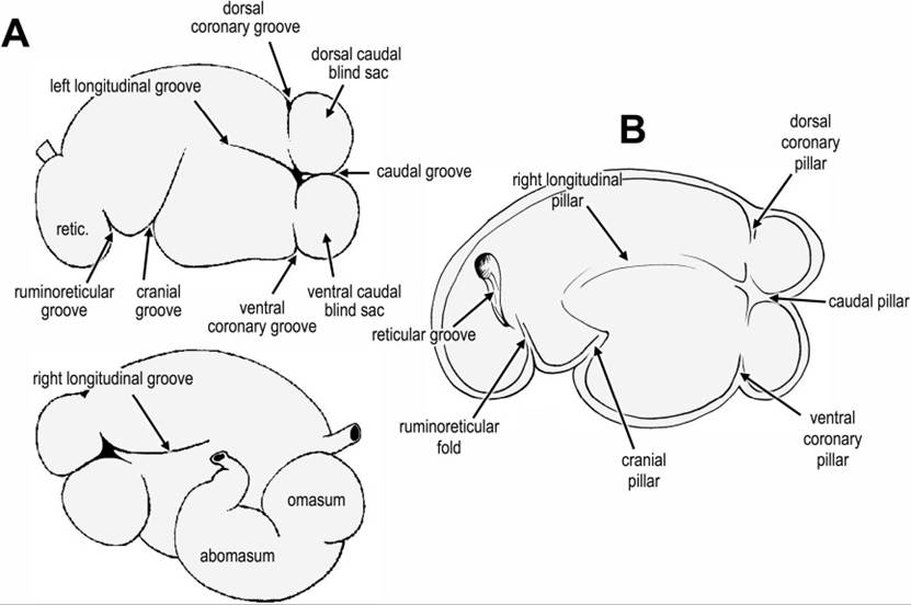
Figure 20-11. Detailed anatomy of the bovine stomach. A) External anatomy from the left (top) and the right (bottom). B) Internal anatomy; grooves on the exterior of the stomach correspond to raised, muscular ridges (“pillars”) on the interior of the stomach.
swallowed typically will fall into and remain in the reticulum; contractions of this part of the forestomach may drive sharp objects through the wall of the stomach, leading to traumatic peritonitis or hardware disease (see Chapter 17). The location of the reticulum immediately caudal to the diaphragm places it opposite the heart, with only the muscular diaphragm between, so that these sharp objects may also be driven into pleural and pericardial spaces.
The reticulum and the rumen (colloquially known as the paunch) are divided ventrally by a thick, muscular ruminoreticular fold. The rumen extends from this fold to the pelvis and almost entirely fills the left side of the abdominal cavity; its capacity depends on the size of the individual but in adult cattle ranges from 110 to 235 L (about 30 to 60 gallons).
The rumen is subdivided internally into compartments by muscular pillars, which correspond to grooves visible on the exterior of the rumen (Fig. 20-10 and 20-11). Right and left longitudinal pillars (corresponding to right and left longitudinal grooves on the exterior) together with cranial and caudal pillars (externally, cranial and caudal grooves) form a nearly complete constricting circle in the horizontal plane.
These divide the rumen into dorsal and ventral sacs. The dorsal sac is the largest compartment. The dorsal sac is continuous crani- ally with the reticulum over the ruminoreticular fold so that the two compartments share a dorsal space.Caudally, the dorsal sac is further subdivided by the dorsal coronary pillars, which form an incomplete circle bounding the dorsal blind sac. The caudal part of the ventral sac forms a diverticulum, the ventral blind sac, separated from the rest of the ventral sac by the ventral coronary pillars.
As in the rest of the forestomach, the mucous membrane lining the rumen is a nonglandular, stratified squamous epithelium. The most ventral parts of both sacs of the rumen contain numerous feathery papillae up to 1 cm long, but papillae are almost entirely absent on the dorsal part of the rumen.
Omasum
The omasum is a spherical organ filled with muscular laminae (an estimated 90-130 in the bovine omasum) that lie in sheets, much like the pages of a book (giving the omasum its colloquial name, book stomach). The stratified squamous mucous membrane covering the laminae is studded with short, blunt papillae. Each lamina contains three layers of muscle, including a central layer continuous with the tunica muscularis of the omasal wall.
The omasum lies to the right of the rumino- reticulum, just caudal to the liver, and in the ox makes contact with the right body wall. The omasum of the sheep and goat is much smaller than the bovine omasum and normally is not in contact with the abdominal wall. Food enters the omasum at the reticulo-omasal orifice, between the laminae, and goes on to the omasoabomasal orifice.
Abomasum
The abomasum (true stomach) is the first glandular portion of the ruminant digestive system (Fig. 20-9). Its proximal portion is ventral to the omasum, and its body extends caudad on the right side of the rumen. The pylorus demarcates the muscular junction of the stomach and small intestine, and like the porcine pylorus, it features an enlarged torus pyloricus.
The epithelium of the abomasum consists primarily of two glandular regions, equivalent to the fundic gland region (region of the proper gastric glands) and the pyloric gland region. The cardiac gland region in the abomasum is confined to a very small area adjacent to the omasoabomasal orifice.