Somatosensation
Pain
Pain is the conscious perception of noxious stimuli. A noxious stimulus is one that is capable of producing tissue damage; it can be thermal, chemical, or mechanical. The receptor for noxious stimuli is the nociceptor, a naked (not encapsulated) nerve ending.
As a rule, axons transmitting noxious information are smaller and less myelinated than those carrying tactile or body position information. Activation of pain fibers of medium diameter and myelination (so-called Aδ fibers) is associated with a sharp, pricking quality of pain as reported by human beings. Activation of the smallest diameter C fibers, which are unmyelinated, produces a dull, burning type of pain. The preponderance of C fibers in visceral sensory fibers explains the burning, aching quality of visceral pain.
As indicated in Chapter 9, a number of ascending spinal cord tracts transmit information about noxious stimuli to brain structures. in addition to projecting to the cerebral cortex for conscious perception, pain pathways typically have strong connections to autonomic centers in the brainstem and parts of the brain that produce increased mental alertness and behavioral and emotional responses to painful stimuli. These connections are responsible for producing signs of sympathetic stimulation (e.g., increased heart and respiratory rates, dilation of pupils), emotional responses, and escape behaviors (Fig. 11-3).
The ability of a given noxious stimulus to produce a perception of pain is a highly mutable property that can be modified in the periphery, in the spinal cord, and in the brainstem.
The threshold of nociceptors in the periphery is not a constant. importantly, many substances released by injured tissues and inflammatory cells stimulate or lower the threshold of nociceptors. Thus, in damaged or inflamed tissue, stimuli that would normally be below threshold for detection may produce activity in nociceptive afferents.
These events contribute to the development of primary hyperalgesia, a phenomenon wherein the perception of pain in injured tissues is increased. A dramatic example of this is made by sunburned human skin; the inflammation of the injured skin lowers threshold of nociceptors so that even a light touch (for instance, contact with clothing) can activate them.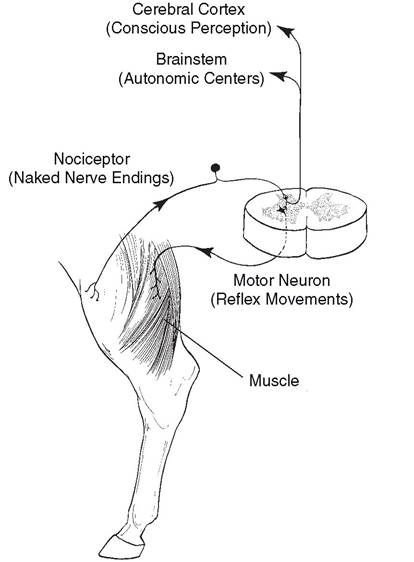
Figure 11-3. Divergence in nociceptive pathways. The primary afferent neuron brings information about painful stimuli into the CNS. From there the information produces reflex movements and goes to the brainstem to produce autonomic and other involuntary responses and to the cerebral cortex for conscious perception.
Events in the dorsal horn of the spinal cord can also affect transmission of nociceptive information. Rapid, prolonged firing of action potentials in the primary afferent neuron can produce changes in the neuron on which it synapses, making it respond more vigorously to subsequent stimulation. This is called windup or spinal facilitation of pain.
Activity in spinal nociceptive pathways is also strongly influenced by descending antinociceptive systems that originate in the brainstem. The midbrain and medulla both possess a series of midline nuclei that inhibit nociception via their connections with nociceptive pathways. These nuclei use multiple neurotransmitters, most notably endorphins, transmitters with powerful antinociceptive properties.
Stoicism, an apparent indifference to pain, is largely determined by personality and training among both humans and animals. High-strung individuals often exhibit exaggerated reactions to stimuli that scarcely merit attention in more “laid-back” individuals. Interestingly, the effectiveness of applying a twitch to a horse’s upper lip as a method of restraint during mildly painful procedures has long been attributed to redirecting the horse’s attention from the procedure to the moderately noxious stimulus of the twitch.
Studies have shown, however, that the correctly applied twitch actually stimulates a release of endorphins, lowers the heart rate, and produces behavioral signs of sedation in the horse. This very old technique of restraint may actually be recruiting some of the neuroanatomic pathways postulated to be stimulated by acupuncture.Proprioception
Proprioception is the nonvisual perception of body position. It is a complex sensory modality that is created through the input of a variety of specialized receptors called proprioceptors. These include joint receptors (providing information on tension and pressure within joints), muscle spindles (signaling changes in muscle length), Golgi tendon organs (signaling tension in tendons), and skin mechanoreceptors (which report contact with the environment).
Ascending proprioceptive pathways project both to the cerebral cortex and the cerebellum. The cerebral cortex uses proprioception to help formulate voluntary motor plans; the cerebellum uses it to adjust ongoing motor movements so that they are smooth and accurate. The information carried in the separate tracts to these targets arise from the same peripheral receptors and primary afferent neurons; it is only the ultimate destination (and therefore use) that is different.
For the cerebral cortex and cerebellum to make effective use of feedback on body position to guide movements, the proprioceptive information must be delivered very rapidly to these brain regions. Consequently, proprioceptive tracts typically have few synapses and are composed of very large diameter, highly myelinated axons (called Aafibers). In fact, the very fastest (up to 120 m/second) axons of the entire nervous system transmit proprioceptive information.
We, and presumably animals, are generally not aware of proprioception, but it is critically important in the execution of accurate, well- coordinated movements. Injury to the proprioceptive pathways results in awkward, inaccurate, uncoordinated gait and movement. The incoordination typical of proprioceptive deficits is referred to as ataxia.
Touch
Touch is the modality associated with non- noxious mechanical contact with the body. Touch receptors are encapsulated, and the axons that transmit touch information to the brain are typically medium in diameter and degree of myelination. Spinal cord tracts associated with touch are found in all the funiculi of the cord.