Species Variations
4.2.1 Horse (Figure 4.1)
The horse's stomach is relatively small, with a capacity of only 5-20 litres depending on the body-size of the horse or pony. It is situated dorsally at the cranial end of the abdominal cavity and never extends to the ventral abdominal wall even when full.
Its parietal surface faces cranially, dorsally and laterally to the left and is in contact with the left lobe of the liver and diaphragm. The visceral surface is related to the left lobe of the pancreas, the diaphragmatic flexure and jejunoileum. The small size of the stomach limits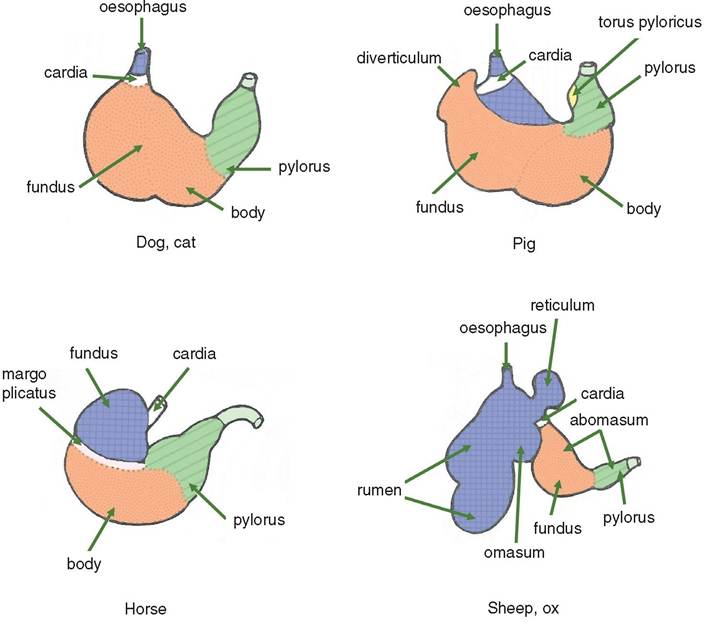
Figure 4.1 The stomachs of the domestic animals. The areas in blue are those parts of the stomach that are lined by a non-glandular, stratified squamous epithelium. The cardiac areas are where the oesophagus enters the stomach in the dog, horse and pig (not shown). In ruminants the cardiac areas are located in the reticulum. In the pig there is a prominent diverticulum of the fundus. In the horse the cardiac zone is very muscular, apparently preventing vomiting. The pyloric zone contains a glandular epithelium (coloured green) and is part of the abomasum in ruminants. The margo plicatus is a distinct ridge separating the glandular (body) and non-glandular areas of the stomach of the horse.
the amount of contents and thereby the quantity of food that can be ingested at any one time. The regions of the stomach are commonly termed as non-glandular (cardia) and glandular (fundic and pyloric). A narrow ridge separating the non-glandular and glandular regions is known as the margo plicatus, and the blind-ending part of the fundic region is termed the saccus caecus.
The relatively short time that ingesta are in the stomach before being passed through the pyloric sphincter leaves little time (20 minutes) for enzymatic activity, resulting in most digestion occurring in the intestines.
No absorption of nutrients occurs in the stomach.Horses are unable to vomit due to several anatomic differences from most other mammals. The sphincter muscle at the oesophageal entry to the stomach is much better developed than in other animals, and their vomiting reflex is virtually non-existent.
4.2.2 Ox (Figures 4.1-4.4)
The stomach of the ox comprises four compartments, three of which are lined by stratified squamous epithelium and termed the rumen, the reticulum and the omasum. The fourth compartment, the abomasum, is lined by a glandular epithelium. The rumen occupies most of the left half of the abdominal cavity. It also extends ventrally and cau- dally onto the right side. Its long axis extends from the level of the ventral part of the seventh intercostal space to the pelvic inlet. Its relations to the omasum and abomasum are shown in Figures 4.2 and 4.5.
Ruminants (cattle, sheep, goats, camelids, deer, giraffes) are unable to produce enzymes to digest cellulose. However, the ruminoreticulum does contain microbes that promote a microfermentation that breaks down the cellulose. This allows access by the enzymes and hydrochloric acid produced by the glandular epithelium of the abomasum to the nutrients locked within the cellulose barrier. The breakdown of cellulose is further aided by rumination. This is the process of sending the contents of the ruminoreticulum for a second mechanical grinding by the teeth. Known as chewing the cud, this practice can occupy the ruminant for up to 8 hours of the day. The omasum is the smallest compartment of the ruminant stomach and is mainly concerned with absorption of water greatly facilitated by the large surface area of the many folds of its inner lining.
The ruminant stomach of the newborn calf is not able to digest the herbivore diet of the adult. In order to digest an early diet of milk, a groove conducts the ingested milk from the cardia directly to the abomasum, bypassing the ruminoreticulum and omasum.
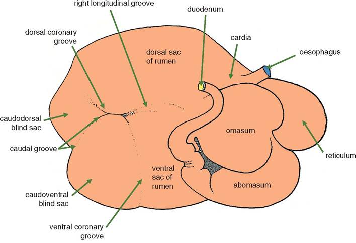
Figure 4.2 Right lateral aspect of the stomach of the ox.
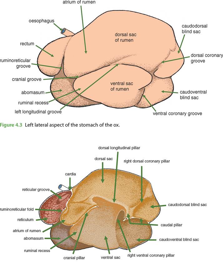
Figure 4.4 The interior of the right half of the rumen and reticulum of the ox.
Muscle fibres in the wall of the groove enable it to close over reflexly and avoid leakage. This important channel is called the reticular groove and is often wrongly called the oesophageal groove.
4.2.3 Sheep (Figure 4.1)
The stomach of the sheep is similar to that of the ox, although the abomasum is relatively larger. The pylorus is adjacent to the ventral 11th or 12th rib on the right.
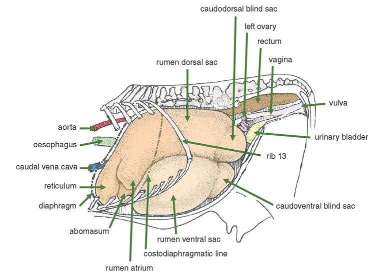
Figure 4.5 Left lateral aspect of the gastrointestinal tract of the ox, in situ. The rumen, as shown, is not completely full and consequently has not extended into the pelvic inlet. The uterine cornu and left ovary are therefore visible.
4.2.4 Pig (Figure 4.1)
The stomach of the pig is relatively large compared with that of the horse. When moderately full it lies entirely within the thoracic cage (Figure 5.4), mainly on the left side. The parietal surface is in contact with the liver and dorsal region of the diaphragm. The visceral surface is related to the spleen on the left and to the coils of the ascending colon (Figure 6.3). When fully distended the stomach makes extensive contact with the abdominal wall, not only ventrally and on the left but on the right side also. Unlike the simple stomachs of the other species, the pig possesses a diverticulum of the fundus that is directed caudally and ventrally. The diverticulum is lined by a glandular mucosa, although its developmental origin is the same as part of the rumen.
4.2.5 Dog
The empty stomach lies entirely within the caudal part of the thoracic cage, in contact with the left region of the visceral (caudal) surface of the liver. It is fixed in position at the cardia by the diaphragm and at the lesser curvature by the lesser omentum. The full stomach extends caudally well beyond the thoracic cage and makes extensive contact with the ventral body wall. In doing so it tends to displace the intestinal mass and the spleen caudodorsally. The stomach is particularly distensible in puppies.
The stomach of the dog is frequently examined by endoscopy for the investigation of vomiting and the presence of foreign bodies. In this procedure the location of the angular incisura in the lesser curvature of the stomach (Figure 4.1) of the dog is an important landmark for endoscopy.
4.3