Species Variations of the Penis
15.5.1 Horse (Figures 15.1, 15.2a and b and 15.3)
The penis of the horse is cylindrical and slightly flattened laterally. It is about 45 cm long and 5 cm in diameter. During erection the penis doubles in length.
The glans penis is the enlarged free end of the penis comprising erectile tissue that is an expanded extension of the corpus spongiosum; it caps the distal end of the corpus cavernosum penis. There are several components of the glans in this species; they are the fossa glandis, collum glandis, corona glandis and urethral process. The paired retractor penis muscle lies ventrally in the midline almost along the whole length of the body of the penis. The corpus spongiosum is covered entirely by the bulbospongiosus muscle. The trabeculae of the corpus cavernosum penis contain bundles of smooth muscle orientated longitudinally (Figure 15.2b). Sympathetic nerves maintain these in a state of tonic contraction and thereby hold the penis within the prepuce. Parasympathetic activity is responsible for erection and also inhibits the sympathetic tonic contraction.Externally there is a strong fibroelastic capsule called the tunica albuginea (see Section 15.3.2 and Figure 15.2b). This type of penis is termed ‘musculocavern- ous' because of the smooth muscle and the large spaces of the corpus cavernosum penis.
The urethral part of the penile body lies ventral to the corpus cavernosum penis and external to the tunica albuginea. In this species the corpus spongiosum is enclosed throughout its length by the bulbospongiosus muscle (Figure 15.2b).
There is an additional arterial supply to the penis in the horse; it is provided by the external pudendal and obturator arteries. There is a substantial drainage of venous
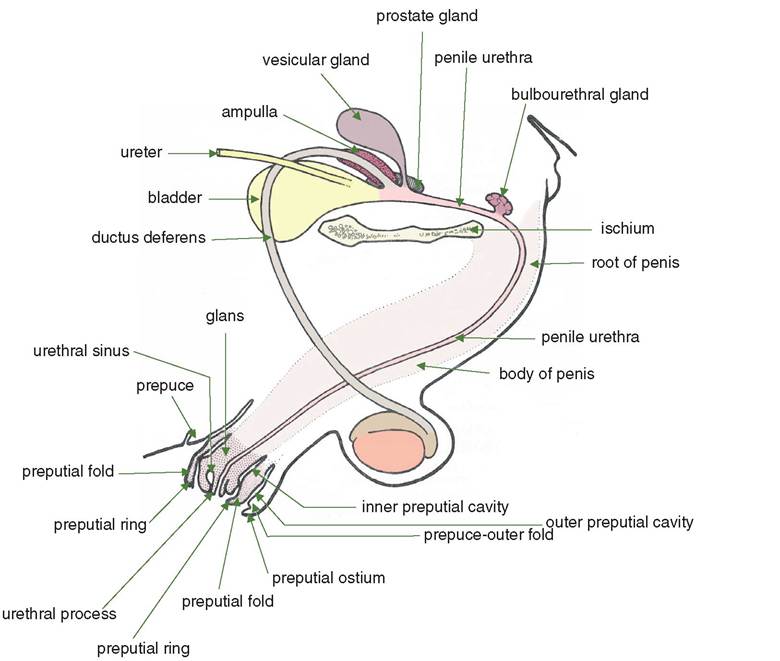
Figure 15.1 Male reproductive organs of the horse.
blood from the penis via the external pudendal vein, which passes through the inguinal canal to enter the external iliac vein. In addition, there is venous drainage via the obturator vein to the internal iliac vein.
15.5.2 Ox (Figures 15.4 and 15.5)
The penis of this species is virtually circular in transverse section. The diameter is smaller than that of the horse (2.5 cm) while its length is much greater (1 m.). When the penis is relaxed there is an S bend, the sigmoid flexure, which is post-scrotal in position (Figure 15.4). The tunica albuginea is reinforced by a strong non-elastic fibrous tissue layer; it encloses the corpus spongiosum as well as the corpus cavernosum. The latter is almost entirely fibrous, with only small areas of erectile tissue remaining. The body of the penis is quite firm but does contain some elastic tissue. Therefore the penis of the ox is classified as ‘fibroelastic. During erection this type of penis hardly shows any increase in diameter. The main effects of the increased blood flow are to straighten out the sigmoid flexure and to harden the glans. The sigmoid flexure returns passively to its
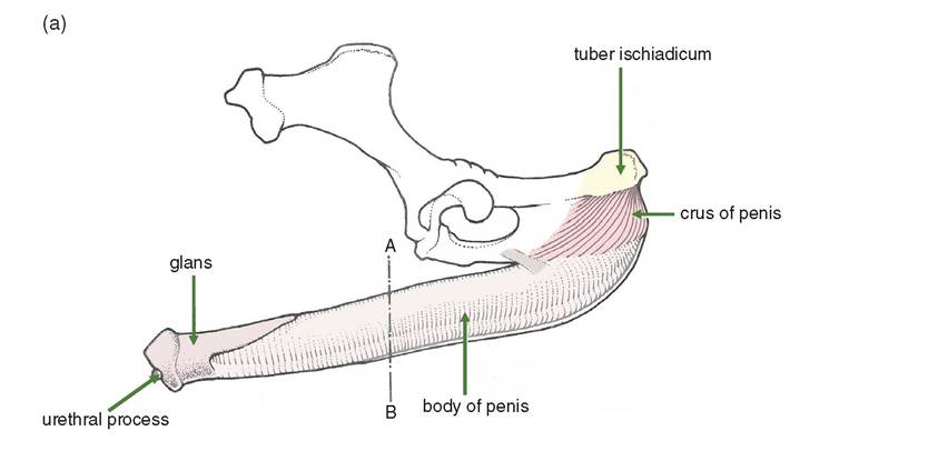
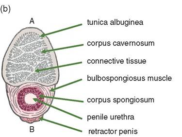
Figure 15.2 (a) Penis of the horse. (b) Transverse section through the body of the penis of the horse at the level A-B in (a).
S shape in the non-erect penis. The retractor penis contributes little to the return of the penis to its non-erect state.
At the tip of the penis the glans penis is an enlarged helmet-like region, which, unlike that of the horse, is not a continuation of the corpus spongiosum. The urethral process is attached to the glans but does not project freely from it.
In the ox there are occasionally abnormal vascular connections that allow leakage from the corpus cavernosum into the corpus spongiosum. In this situation erection fails to occur.
15.5.3 Sheep (Figure 15.6)
The crura and body of the penis are similar to those of the ox. The glans penis is relatively larger than that of the ox. The urethral process is a worm-like extension of the
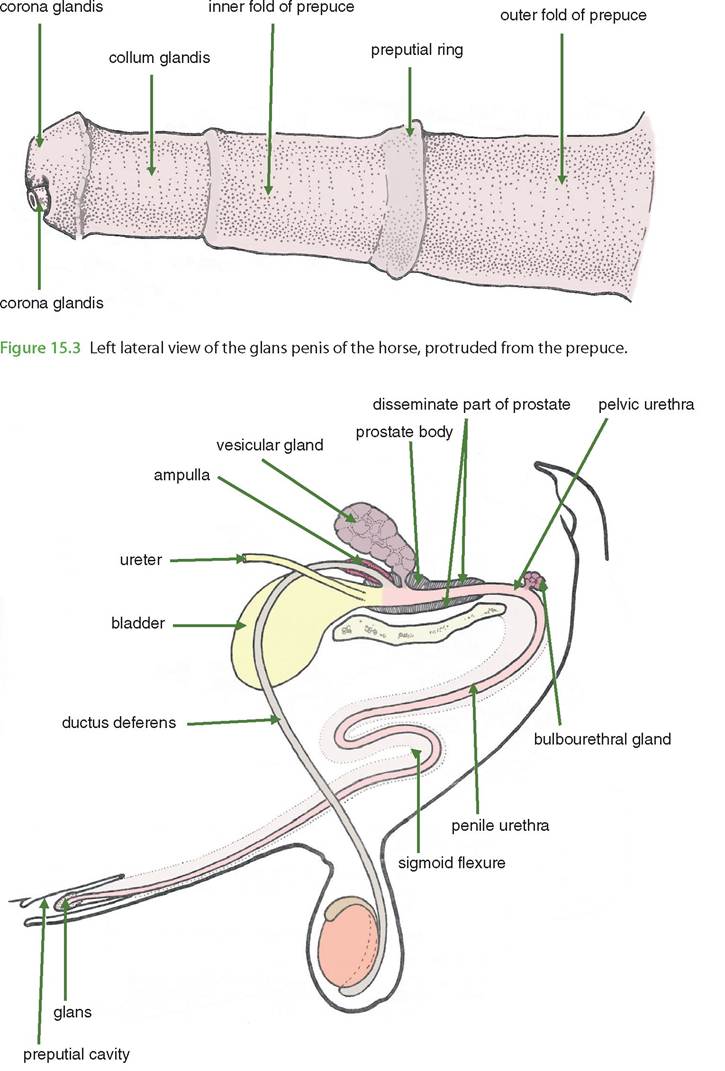
Figure 15.4 Male reproductive organs of the ox. The outline of the body of the penis is indicated by dotted lines.
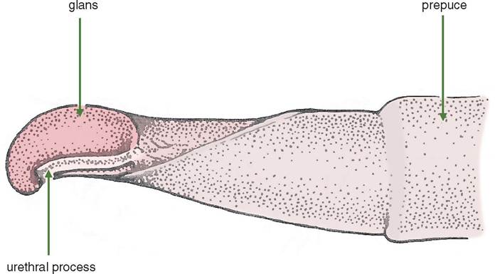
Figure 15.5 Left lateral view of the penis of the ox, protruded from the prepuce.
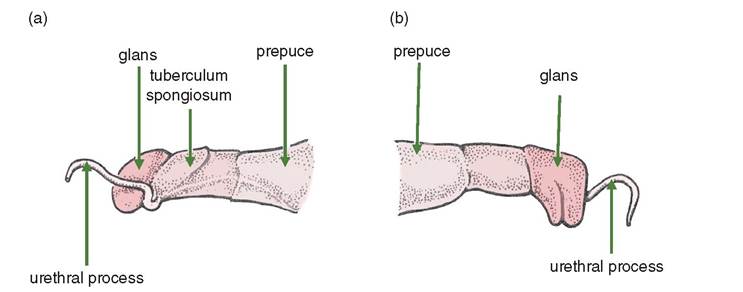
Figure 15.6 The free part of the penis of the sheep, protruded from the prepuce. (a) Left lateral view and (b) right lateral view.
urethra beyond the glans; it is about 2 cm in length. Uroliths can cause an obstruction to urethral function and require surgical removal or amputation of the process. The tuberculum spongiosum is an asymmetrical rounded projection of the corpus spongiosum penis on the left lateral aspect of the free part of the penis.
15.5.4 Pig (Figures 15.7 and 15.8)
The penis of this species is similar to that of the ox in that it is fibroelastic and comprises two crura and a body. However, the sigmoid flexure is prescrotal since the scrotum is virtually subanal in position. There is no glans penis in the pig, the free part being spirally twisted, slightly flattened and ending in a point. The urethral orifice is a slit-like
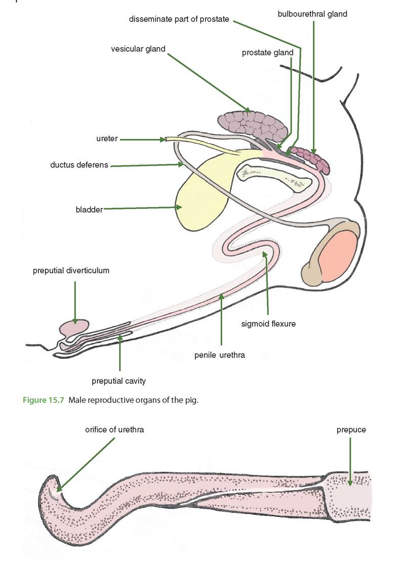
Figure 15.8 Ventral view of the free part of the penis of the pig, protruded from the prepuce.
opening near the pointed tip. There is no urethral process as in the ruminant. The pig squirts urine rather than as a continuous stream.
The boar can ejaculate up to 400 ml and take 5-10 minutes to do it.
15.5.5 Dog (Figures 15.9 and 15.10)
This species demonstrates several structural differences as compared with the other species.
1) Whereas there are paired crura that fuse as the body of the penis, in the caudal half of the penis the left and right corpora cavernosa penis remain separated by a midline septum.
2) The cranial part of the corpora cavernosum penis is completely ossified and has no erectile tissue; it is called the os penis. The urethra, enclosed in the corpus spongiosum, fits in a ventral groove in the os penis. The os penis ends just before the cranial end of the non-erect glans penis. The corpus cavernosum does not have a significant role in erection, but the erectile tissue is supplied with blood by the pudendal artery and drains via the internal pudendal vein.
3) The corpus spongiosum penis also differs from that in the other species. There are two large expansions of the corpus spongiosum: (i) the bulbus glandis located at the level of the caudal os penis, and (ii) the bulb of the penis located between the crura. It is the bulbus glandis that plays an important role during intromission.
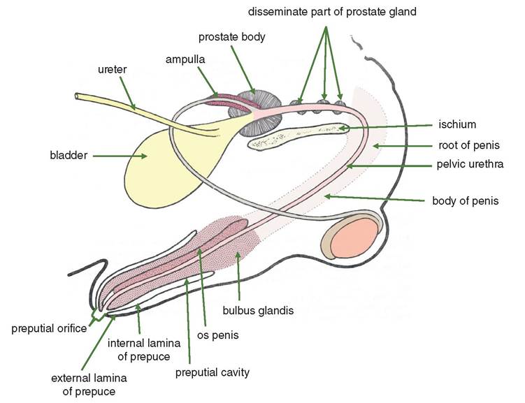
Figure 15.9 Male reproductive organs of the dog.
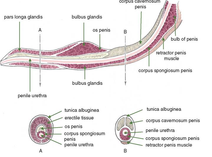
Figure 15.10 Penis of the dog with transverse sections through the glans at A and through the body of the penis at B.
The glans penis of the dog consists of the bulbus glandis and the pars longa glandis. The latter is the most distal (cranial) part of the corpus spongiosum, extending from the bulbus glandis to the tip of the penis. During erection the pars longa glandis increases in length.
Intromission is possible before complete erection is achieved through the stiffness of the penis conferred by the os penis. Complete erection occurs after intromission and involves enlargement of the bulbus glandis within the vestibule of the bitch. Once this is achieved it is not possible to withdraw the penis for 5-45 minutes after ejaculation.
15.5.6 Cat (Figure 15.11)
The penis of this species is similar to that of the dog apart from the following features.
The non-erect penis points caudally (instead of cranially as in the dog). The preputial orifice is ventral to the scrotum, which is subanal. During erection the penis pivots at its root so that the glans becomes directed cranially.
There is an os penis, but it is quite short. The glans is short and studded with small cornified barbs that serve to hold the penis firmly in the vestibule of the female.
15.6