Stay Apparatus
Ligamentous structures that store and release energy during locomotion are also found proximal to the foot. These elements of the stay apparatus are also the elements of the limb that permit the horse to stand with minimal physical effort.
To understand the function of the stay apparatus, it is necessary to keep several concepts central: (1) Ligaments, while elastic, do not stretch nearly as much as muscle. Therefore tendons, because they connect with muscles, offer much less resistance to stretch than do ligaments. (2) When joints are loaded (i.e., bearing weight), they tend to collapse. (3) To keep joints from collapsing with minimal muscular effort, ligamentous straps must cross the joint to counteract the tendency to fold during weight bearing (Fig 14-12).
Thoracic Limb
The thoracic limb is in part affixed to the trunk by the fan-shaped m. serratus ventralis, extending from the scapula to the ribs. This muscle is characterized by a thick, tendinous layer that is capable of supporting the trunk without contraction of the muscle fibers. The weight of the trunk is thereby supported effortlessly by a slinglike structure composed of right and left mm. serrati ventrales (see Fig. 7-7); by this construct, much of the weight of the horse is transferred to the thoracic limbs.
When bearing weight, the shoulder joint tends to flex, and the fetlock, pastern, and coffin joints tend to hyperextend, while the carpus and elbow are relatively stable when loaded in the extended position. Components of the stay apparatus cross all of these joints,
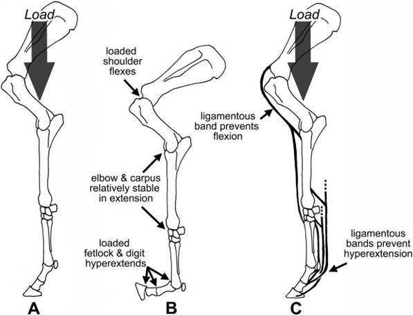
Figure 14-12. When the appendicular skeleton is loaded with weight (A), the joints tend to collapse (B). The stay apparatus is a series of ligamentous bands that cross the joints and passively prevent this collapse (C).
counteracting their tendency to collapse under load (Fig. 14-13).
The extensor surface of the shoulder is crossed by the tendon that is the origin of the m. biceps brachii, a very broad, partly cartilaginous tendon that is continuous with a very dense fibrous band running through the length of the muscle belly. This fibrous band is continuous with the tendons of insertion, including the lacertus fibrosus, a substantial band that blends into the deep fascia (epimysium) of the m. extensor carpi radialis. This feature of the m. biceps brachii creates a continuous ligamentous connection from the scapula through the length of the brachium, across the elbow, and to the proximal metacarpus via its connection with the tendon of the m. extensor carpi radialis. it is this band of connective tissue that will, without muscular effort, counteract flexion of the shoulder when the limb is bearing weight. This same continuous band, through its connection with the m. extensor carpi radialis, contributes further to the stability of the carpus in extension.
The elbow is surprisingly stable when fully extended and loaded. The m. triceps brachii, the primary extensor of the elbow, contributes to this stability by maintaining a slight amount of tone, even when the horse is sleeping on his feet.
The rest of the stay apparatus is primarily concerned with holding the fetlock and to a lesser extent pastern and coffin joints in a physiologic position. Without a ligamentous check on the palmar aspect of these joints, the fetlock would drop to the ground and the toe would point upward, with the sole off the ground when the limb was loaded.
The primary support of the fetlock and pastern joints comprises the suspensory ligament, the proximal sesamoid bones, and the ligaments of the proximal sesamoid bones. Recall that these structures form a continuous ligamentous connection between the palmar aspect of the carpus and proximal metacarpus distal to the proximal and middle phalanges.
The tendons of both digital flexor muscles offer additional support to the fetlock and pastern joints, and the tendon of the deep digital flexor muscle resists hyperextension of the coffin joint. Remember, though, that muscle tends to stretch easily. These digital flexors can support the distal joints without muscular effort
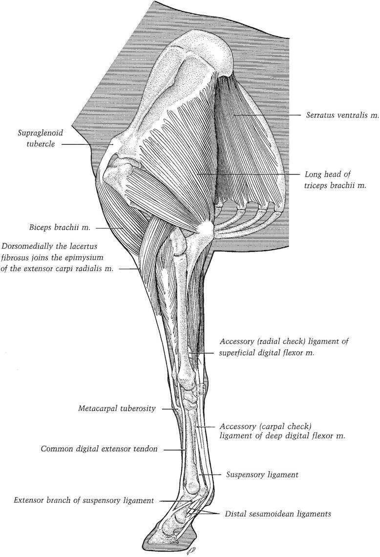
Figure 14-13. Stay apparatus of the thoracic limb. (Reprinted with permission of Wiley-Blackwell from Stashak
T.S. Adams’ Lameness in Horses. 5th ed. Baltimore: Lippincott Williams & Wilkins, 2002.)
because they both feature accessory ligaments, ligamentous connections between more proximal bones and the insertions of the tendons.
The superficial digital flexor muscle possesses a ligamentous head that attaches to the caudal aspect of the distal radius and joins the muscle’s tendon near the carpus. This accessory ligament of the superficial digital flexor muscle is commonly known as the radial or proximal check ligament, and its presence creates a continuous, ligamentous band from the radius to the insertion of the tendon on the proximal and middle phalanges. This provides additional support to fetlock and pastern.
The deep digital flexor muscle also features an accessory ligament, this one attaching to the caudal part of the carpal joint capsule and blending with the muscle’s tendon just distal to the carpus. This accessory ligament of the deep digital fl exor muscle is more commonly called the carpal or distal check ligament. it creates a continuous ligamentous band that extends from the carpus to the distal phalanx, supporting all of the joints in the digit.
Injury to the flexor tendons produces some dropping of the fetlock as some of the resistance to hyperextension is lost. If injury includes the deep digital flexor tendon, the toe is likely to come off the ground, as this tendon is the only one resisting hyperextension of the coffin joint. The most devastating injuries, however, involve the suspensory ligament and/or the proximal sesamoid bones.
Fractures of the proximal sesamoids are the most common of all fractures in the forelimb. If the fractures are complete (transverse fractures of both sesamoids), or if the suspensory ligament is transected, the ligament loses its connections to the phalanges and the fetlock will drop to the ground. Such an injury is often irreparable and may necessitate destruction of the horse.Pelvic Limb
Distal to the tarsus, the stay apparatus in the pelvic limb is more or less identical to that of the thoracic limb (Fig. 14-14). The accessory ligament of the deep digital fl exor muscle (tarsal check ligament) arises from the plantar aspect of the tarsus and proximal metatarsus; this accessory ligament is rather long and very slender and may even be absent in some individuals. The superficial digital flexor muscle of the pelvic limb is nearly entirely tendinous and therefore needs no accessory ligament to create a continuous band from origin (caudal distal femur) to insertion on proximal and middle phalanges. Furthermore, the tendon of the superficial digital flexor muscle inserts on the tuber calcanei, providing, in effect, a check ligament at this point.
For the pelvic limb to support weight without collapsing, the stifle and hock must be prevented from flexing. This is accomplished by one mechanism to lock the stifle in extension and a second mechanism (the reciprocal apparatus) that guarantees that the hock will always flex and extend in unison with the stifle. The reciprocal apparatus provides that as long as the stifle is locked in extension, the hock will be likewise.
Recall that the equine patella features a hook-shaped medial parapatellar cartilage and three patellar tendons that insert on the tibial tuberosity (Fig. 6-8). During movement, the patella glides up and down the femoral trochlea as the quadriceps muscle contracts and relaxes, respectively. When the horse stands at rest, however, the patella is drawn proximad so that the parapatellar cartilage and medial patellar tendon are hooked over the large medial ridge of the trochlea.
in this position, the stifle is held in extension with minimal muscular effort. Typically, the horse activates this mechanism in one pelvic limb, resting the other with the toe on the ground; this posture is sometimes referred to as standing “hip-shot.”The hock and stifle normally flex and extend in unison because of the reciprocal apparatus, a combination of ligamentous structures connecting the femur with the tarsus and metatarsus (Fig. 14-15). On the cranial aspect of the crus, the peroneus tertius, an entirely ligamentous structure, extends from the region of the lateral femoral condyle to insertions on the tarsus and proximal metatarsus. in doing so, it
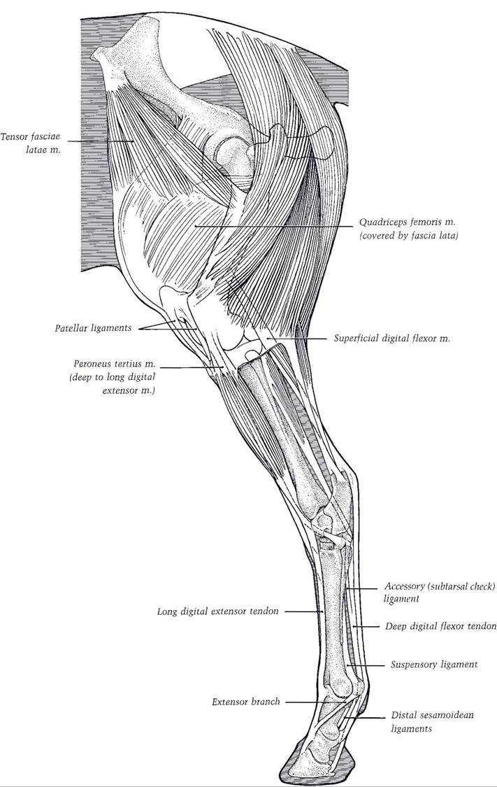
Figure 14-14. Stay apparatus of the pelvic limb. (Reprinted with permission of Wiley-Blackwell from Stashak
T.S. Adams' Lameness in Horses. 5th ed. Baltimore: Lippincott Williams & Wilkins, 2002.)
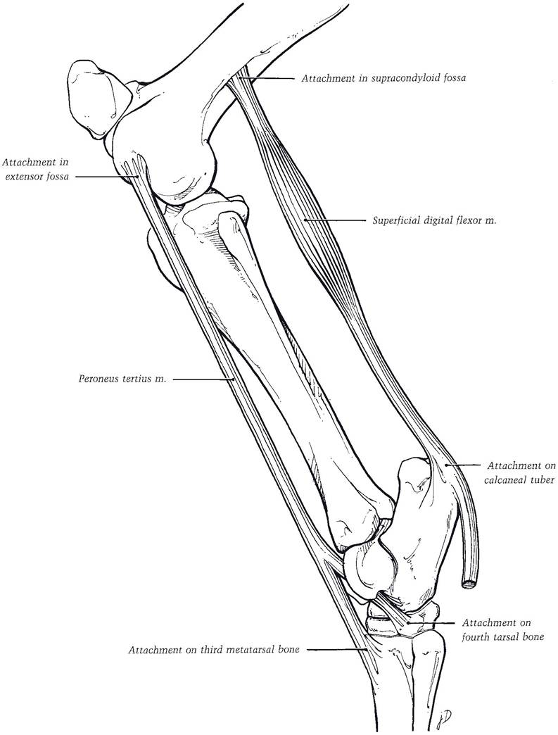
Figure 14-15. Reciprocal apparatus. (Reprinted with permission of Wiley-Blackwell from Stashak T.S. Adams’
Lameness in Horses. 5th ed. Baltimore: Williams & Wilkins, 2002.)
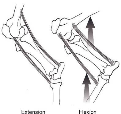
Figure 14-16. The change in the angle in the stifle is accompanied by a similar change in the hock.
forms a relatively Unstretchable band across the extensor surface of the stifle and the flexor surface of the tarsus. on the caudal aspect of the crus, the superficial digital flexor muscle, which is also entirely tendinous, arises from the distocaudal femur and inserts on the calcaneus and digit. This forms a ligamentous connection that crosses the flexor surface of the stifle and extensor surface of the tarsus. As shown in Figure 14-16, these elements create a parallelogram of ligaments, wherein the change in angle in the stifle is accompanied by a similar change in the tarsus.
A horse standing with the stifle in a locked position necessarily has locked the hock in extension as well.In Upwardfixation of the patella, the patella locks above the medial ridge of the femoral trochlea at inappropriate times. The condition may be due to conformational abnormalities or a neuromuscular disorder. The affected animal exhibits a sudden extension of stifle (and hock) that may be intermittent or persistent. Conservative treatment involves conditioning exercise to improve the horse’s ability to control the patella. If this fails, the medial patellar ligament can be transected; this prevents the patella from becoming fixed over the ridge.
Can horses sleep standing up? Yes and no. Sleep is divided into five stages, numbered I through V. Stages I through IV are a continuum of increasing depth of sleep, with I and II indicating light sleep (drowsing) and III and IV indicating deep sleep. Stage V is what is described in humans as REM (rapid eye movement) sleep. This is the part of sleep most usually associated with dreaming. One of the defining characteristics of REM sleep is that other than eye and respiratory muscles, all skeletal muscles are in a state of flaccid paralysis. Horses are awake about 19 hours of a day and spend about 2 hours each in light and slow-wave sleep, during which they can stand with only the most minimal of muscle tone, thanks to the stay apparatus. Horses normally have REM sleep for only about an hour a day, but because REM sleep is associated with paralysis of voluntary muscles, horses must lie down during that time.