STRUCTURE
The skin is thick over the flank but thins ventrally, particularly in heavy draft animals. It is especially thin in the cleft between the abdomen and thigh where it is sparsely haired and glistens with the secretion of the sebaceous glands concentrated here.
In contrast, sweat glands are most abundant over the flank.A large subcutaneous bursa, a postnatal development, is present over the coxal tuber. Elsewhere the skin is closely adherent to the cutaneous trunci, which cover most of the flank, though not the abdominal floor. The upper border of the cutaneous muscle follows a line drawn from the withers to the stifle. The muscle is thickest cranially where it extends into the fascia over both the lateral and the medial aspects of the shoulder and arm. Caudally, it continues within the flank fold to end on the lateral femoral fascia. The cutaneous muscle is employed to twitch the skin to dislodge flies and other irritants. No detached bundles are associated with the prepuce, as in many species.
The loose fascia deep to the muscle conveys the cutaneous nerves and superficial vessels and encloses the subiliac lymph nodes.
The deeper fascia consists largely of elastic tissue and, being yellowish, is also known as the tunica flava. It is well adapted to the passive support of the viscera and is thickest ventrally, where the burden is greatest. The dorsal part is easily dissected from the underlying muscle, but its ventral part exchanges fibers with the
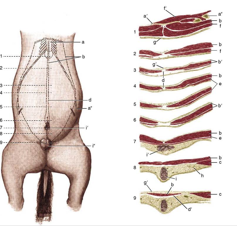
Figure 21-1 Changes in the structure of the abdominal floor shown by means of a series of transverse sections (1-9) of a gelding. a, Sternum; a', xiphoid cartilage; a", costal arch; b, rectus abdominis; b', rectus sheath; c, internal oblique; d, linea alba; d', prepubic tendon; e, cutaneous trunci; f, pectoralis ascendens; f, diaphragm; g, skin; g', fat; h, superficial inguinal lymph nodes; i, penis; i', prepuce; i" scrotum.
aponeurosis of the external oblique and is more tightly adherent. Bands detached from the tunic help support the prepuce or the udder. Careful suturing of this layer is necessary after abdominal surgery because its elastic nature tends to evert and draw apart the edges of a wound in the underlying muscle.
Before considering the muscles of the abdominal wall, it is necessary to pay attention to the linea alba and prepubic tendon since these and the associated structures have a particular importance in the horse. The linea alba, mainly formed from the aponeuroses of the flank muscles, is considerably strengthened by longitudinal fibers. It is unequally developed along its length, being widest where it carries the umbilical scar (see Figure 21-1/d). It finally combines with the insertion tendons of the right and left rectus abdominis muscles to form a broad plate.
This may be regarded as the initial formation of the prepubic tendon through which the abdominal muscles find attachment to the pelvic skeleton (Figure 21-2/5). Once formed, the tendon ascends almost vertically
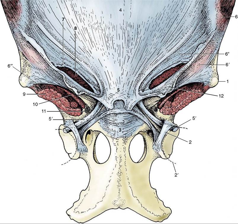
Figure 21-2 The attachment of the abdominal muscles on the pelvis and the prepubic tendon. 1, Coxal tuber; 2, transverse acetabular ligament; 2', femoral head; 3, pubis; 4, tunica flava over linea alba; 5, prepubic tendon; 5', accessory ligament; 6, external abdominal oblique; 6', 6", pelvic and abdominal tendons of external oblique aponeurosis; 6'", attachment of pelvic tendon of external oblique aponeurosis on sartorius and iliopsoas (“inguinal ligament”); 7, superficial inguinal ring; 8, internal abdominal oblique; 9, iliopsoas; 10, sartorius; 11, vascular lacuna containing femoral vessels; 12, femoral fascia (lamina).
toward the pelvic brim, but before reaching this, it is augmented by a strong transverse thickening. This thickening is mainly formed by the tendons of origin of the pectineus muscles (of the thighs), which arise from both the ipsilateral and contralateral pubic bones (from and medial to the iliopubic eminences) and which thus partly decussate across the midline.
Additional but lesser contributions to the prepubic tendon are made by the caudal margins of the oblique abdominal muscles and the cranial part of the gracilis. A feature of great interest, peculiar to the horse, is the detachment from the caudolateral aspects of the prepubic tendon of the stout rounded cords that furnish accessory ligaments to the hip joints (Figure 21-2/5' and Figure 21-1). Each accessory ligament crosses the ventral surface of the pubis, heading toward the acetabulum, which it enters through the notch in the rim; it ends by inserting on the head of the femur beside the intracapsular ligament (of the head of the femur) that is found in all species. Each accessory ligament is predominantly composed of fibers from the two rectus muscles, and many fibers have decussated from the contralateral side. The ligaments appear to be the principal insertions of these muscles. Their existence partly explains the restrictions on the movements permitted at the equine hips. It is postulated that the accessory ligaments are tensed by the weight of the abdominal contents and that this tension helps secure the femoral heads in place.Since the main weight of the abdominal organs is carried by the prepubic tendon, it follows that its rupture
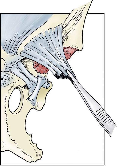
Figure 21-3 The origin of the external spermatic fascia and femoral lamina from the margin of the superficial inguinal ring. (See Figure 21-2 for orientation.)
has the most dire consequences. This mishap, fortunately rare, is for obvious reasons most common in heavily pregnant mares.
The external abdominal oblique (Figure 21-4/7) is the most extensive muscle of the flank. It arises from the thoracolumbar fascia and also from the lateral aspect of the thoracic wall (from the fifth rib caudally) by a series of digitations that engage with those of the serratus ventralis. The majority of its fascicles run caudo- ventrally to a broad aponeurosis that succeeds the fleshy part of the muscle along a line that sweeps from the coxal tuber toward the ventral end of the fifth rib.
Before insertion, the aponeurosis splits into (1) a large abdominal tendon that continues over the rectus to reach and insert on the linea alba and (2) a small pelvic tendon that inserts on the coxal tuber, the fascia over the iliopsoas and sartorius muscles, and the prepubic tendon (see Figure 21-2). The split between the two tendons constitutes the superficial ring of the inguinal canal (Figure 21-4/5). (The margins of the
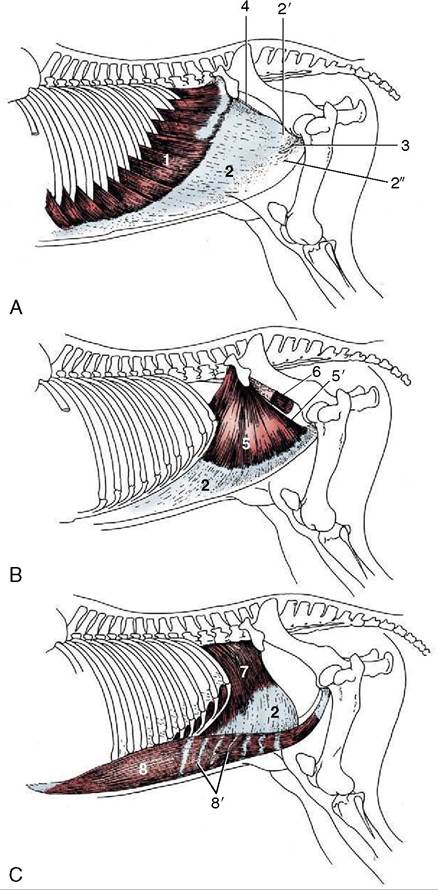
Figure 21-4 The abdominal muscles and their skeletal attachments. 1, External abdominal oblique, muscular part; 2, aponeurotic parts of 1, 5, and 7;2', 2'', pelvic and abdominal tendons of aponeurotic part; 3, superficial inguinal ring; 4, attachment of pelvic tendon of external oblique aponeurosis on iliopsoas and sartorius (“inguinal ligament”); 5, internal abdominal oblique, muscular part; 5', free caudal border forming the cranial margin of the deep inguinal ring; 6, iliopsoas, partly enclosed by iliac fascia; 7, transversus abdominis, muscular part; 8, rectus abdominis; 8', tendinous inscriptions.
tendons are known as crura where they bound the opening, but the term is often misapplied to the tendons themselves.)
The unnecessary term inguinal ligament confuses many descriptions of these structures. It is sometimes specifically applied to the thickened caudodorsal edge of the pelvic tendon. In fact, the prominence of this edge (Figure 21—4/4) owes less to thickening than to tension through its connection with the fascia covering the iliopsoas and sartorius.
The internal oblique muscle (Figure 21-4/5) radiates from an origin concentrated on the coxal tuber but extending onto the dorsocaudal edge of the pelvic tendon of the external oblique. Most bundles run cra- nioventrally to insert on the last costal cartilages or, via an aponeurosis that fuses with that of the external oblique, into the linea alba. Some pass ventrally and caudoventrally, and these cover the superficial inguinal ring on its internal aspect (Figure 21-5/4).
A caudal slip provides the cremaster, which passes onto the spermatic cord. The junction of the fleshy and aponeurotic parts of this muscle occurs more than halfway down the abdominal wall.The transversus abdominis (Figure 21—4/7) takes origin from the lumbar vertebrae and the medial aspect of the last ribs, ventral to the origin of the diaphragm.
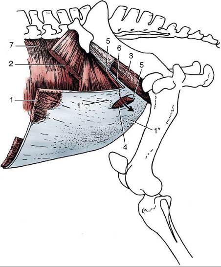
Figure 21-5 The muscles of the inguinal region. The arrow passes through the inguinal canal. 1, External abdominal oblique; 1', 1", pelvic and abdominal tendons of external oblique aponeurosis; 2, internal abdominal oblique; 3, iliopsoas partly enclosed by iliac fascia; 4, superficial inguinal ring; 5, cranial border of deep inguinal ring; 6, attachment of pelvic tendon of external oblique aponeurosis on iliopsoas and sartorius (“inguinal ligament”); 7, transversus abdominis.
The fleshy part is continued by an aponeurosis that passes deep to the rectus abdominis to reach the linea alba. The transversus, the least extensive of the three muscles of the flank, does not extend caudal to the level of the coxal tuber; the internal lamina of the rectus sheath is thus deficient caudally.
The rectus abdominis (Figure 21—4/and femoral lamina, which appear to continue the lateral crus directly (see Figure 21-3). The medial (ventral) crus is somewhat frayed but can be identified on palpation through the skin. This is best performed by placing the palm against the belly and advancing the fingers into the cleft between the thigh and abdominal wall. The lateral crus is passed unnoticed, but the medial crus is recognized as a firm edge. The fingers pass into the outer part of the canal most readily with the thigh abducted (when the femoral fascia [lamina] draws the lateral crus outward). It follows from the orientation of the deep and superficial rings that the canal has a triangular outline; it is relatively long cranially and very short caudally where the two openings butt against the prepubic tendon (see Figure 21-5).
The peritoneal sheath (vaginal tunic) of the spermatic cord contains a cavity that places the space about the testis in free communication with the peritoneal cavity of the abdomen. The communication occurs through the vaginal ring (ca. 3 cm long) situated midway in the deep inguinal ring (see Figure 22-19/10 and Figure 22-24, A-B); the vaginal ring, with the constituents of the spermatic cord converging on it, can be identified per rectum in the stallion. The vaginal cavity provides a possible route for the herniation of intestines that may even reach the scrotum. This occurrence (indirect inguinal hernia) is a comparatively common sequel to castration. Direct inguinal hernia, in which a loop of intestine forces an entry into the canal beside the vaginal tunic, is rare in horses.
Incomplete descent of one or both testes (cryptorchidism) is common in the horse (p. 579). The testis may be retained within the abdomen or may enter but fail to leave the canal. Surgical correction may be indicated. It is therefore necessary to be aware that while the spermatic cord occupies a central position within the canal, the external pudendal artery, which must be treated with respect, occupies the caudomedial corner. The artery is accompanied by the genitofemoral nerve and a small vein; the larger (accessory) external pudendal vein makes a separate passage between the pectineus and gracilis muscles.