STRUCTURE
The ventrolateral wall of the abdomen is composed of as many as 9 or 10 layers, although not all cover the entire extent. The skin is freely movable except over the coxal tuber. The cutaneous muscle is thick over the lower parts of the flank but thins dorsally and does not extend over the paralumbar fossa; it also leaves the abdominal floor bare except for detached fascicles that supply the male animal with cranial and caudal muscles of the prepuce.
The cutaneous muscle extends through the flank fold to end in an aponeurosis over the lateral surface of the thigh (Figure 28-1, A).The loose superficial fascia provides pathways for the cutaneous nerves and encloses certain lymph nodes. The elongated subiliac node lies vertically within the skin fold, pressed against the cranial margin of the thigh some distance above the patella; it can always be found on palpation. It drains the more superficial layers of the body wall as far forward as the caudal part of the thorax and also receives lymph coming from the skin and superficial muscles of the thigh and croup (see Figure 29-46). A number of smaller nodes within the paralumbar fossa drain the surrounding parts; they normally escape notice but appear as circumscribed swellings when enlarged. The subcutaneous abdominal (“milk”) vein runs forward over the abdominal floor from the udder (see Figures 29-34 and 29-44).
The deep fascia is transformed into an elastic tunica flava, attached to the underlying muscle and sharing in supporting the viscera. Ventrally it gives origin to the external spermatic fascia or the medial lamina of the suspensory apparatus of the udder.
The muscle layer is broadly arranged as in other species. In the flank it consists of a triple layer of flat muscles that take origin from the ribs, lumbar transverse processes, and ilium (see Figure 28-1). These are continued over the abdominal floor by aponeurotic tendons that enclose the rectus muscles to each side of the linea alba where the aponeuroses attach (see Figure 1-37).
The linea alba runs from the xiphoid process of the sternum to the center of the prepubic tendon, where it blends with the end tendons of the recti.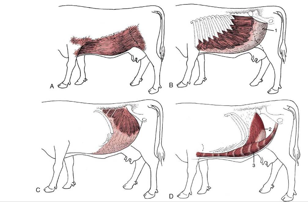
Figure 28-1 Cutaneous trunci and abdominal muscles. A, Cutaneous trunci, especially well-developed ventrally. B, External abdominal oblique with superficial inguinal ring (1) in its aponeurosis. C, Internal abdominal oblique. D, Transversus abdominis (2) and rectus abdominis (3). Note the reduction in the thickness of the wall along the caudal part of the rectus margin.
The most superficial muscle of the flank, the external oblique, arises by fleshy serrations from the outer surfaces of the last eight ribs. Its most dorsal fibers run more or less horizontally toward the coxal tuber, but the greater number slope caudoventrally to find attachment to the linea alba (Figure 28-1, B). The gap that intervenes between the dorsal border and transverse processes is closed by a sheet of fascia. The fleshy part is succeeded by an aponeurotic tendon, and the transformation occurs along a line that first drops vertically, from a point roughly level with the coxal tuber, before sweeping cranially. A split within the aponeurosis provides the superficial opening (ring) of the inguinal canal.
The second muscle, the internal oblique, has a tendinous origin from the coxal tuber and the pelvic tendon of the external oblique and several independent fleshy origins from the tips of the lumbar transverse processes. It radiates to insert on the last rib and into the linea alba. Most fibers run cranioventrally, but the thicker, most caudal fascicles pass slightly behind the plane of the tuber. The muscle-tendon junction slopes caudo- ventrally, and only the most caudal strip is fleshy where the muscle crosses the margin of the rectus (Figure 28-1, C). The aponeuroses of the two oblique muscles become increasingly interwoven where they pass ventral to the rectus and together furnish the external layer of the rectus sheath.
The flesh of the internal oblique forms the inner wall of the inguinal canal.The third, the transversus abdominis, arises from the last ribs and the extremities of the lumbar transverse processes. Its craniodorsal triangle is tendinous, but most of the part covering the flank is fleshy; before reaching the edge of the rectus, the flesh gives way to an aponeurosis that crosses the dorsal face of the rectus to gain the linea alba, thus forming the inner layer of the rectus sheath. Most fibers run transversely, and none pass behind the plane of the coxal tuber; the dorsal surface of the rectus is thus left uncovered in its most caudal part (Figure 28-1, D).
The rectus abdominis muscle is interrupted in the usual way by several tendinous intersections (Figure 28-1/5). It arises from the outer surfaces of the lower ends of the last 10 ribs and continues as a wide band separated from its neighbor by the flattened linea alba; it narrows suddenly as it approaches the pubic brim, and the tendon that succeeds the flesh twists to form with its fellow and the linea alba a V-shaped trough that continues as the central part of the prepubic tendon. Before reaching the pubic brim, which it approaches almost vertically from below, the prepubic tendon is strengthened by joining the decussation formed by the contralateral parts of the pectineus muscles (each of which arises from both pubic bones) and by additional contributions from the aponeuroses of the abdominal oblique muscles. Ultimately, and after partial decussation, the rectus tendons end in common on the symphysial crest of the pelvis and on the medial symphysial tendon that arises here. A rounded median depression of the internal surface of the prepubic tendon is ascribed to the drag of the udder (see Figure 29-40).
A thin fascia covers the abdominal muscles internally and supports the parietal peritoneum. The largest deposits of fat in the subperitoneal tissues are encountered toward the pelvic inlet. The wholly tendinous nature of a region of the abdominal wall, along the border of the rectus in front of the stifle, merits emphasis.
The inguinal canal resembles that of the horse (p. 549) so closely that a separate description is unnecessary. Inguinal hernias are infrequent in cattle but common in male sheep, although there are no obvious differences in the adult anatomy. It is probable that the frequent incidence in rams is connected with inherited anomalies in gubernacular development.