STRUCTURE OF THE NERVOUS SYSTEM
1. Differentiate between dendrites and axons.
2. Sketch a multipolar neuron.
3. What is a nerve fiber?
4. What is the difference between a neurilemma and the axolemma?
5.
What is a nucleus?6. What is a ganglion?
7. What is a tract?
8. What is a nerve?
9. What are the components of a synapse?
0. What are characteristics of a synapse?
11. Which one of the glial cells facilitates transport of blood constituents from capillaries to neurons?
2. Are myelin sheaths in the central nervous system and peripheral nervous system formed by the.same cells?
3. How do myelin sheaths of the central nervous system and peripheral nervous system differ?
4. At what location does the axolemma have contact with extracellular fluid in myelinated nerve fibers?
The many complex functions of the nervous system are accomplished by two cell types: neurons and glial cells, also called neuroglia and glia. Neurons transmit nerve impulses and join with others via synapses. Glial cells provide service to neurons and their environment. The human brain contains approximately 100 billion neurons and a 10-fold number more of glial cells.
Neurons and Synapses
The neuron (nerve cell) consists of the cell body and all its processes, the dendrites, and the axon (Figure 4-1). A nerve cell process is a dendrite if it conducts impulses toward the cell body and it is an axon if it conducts impulses away from the cell body. A neuron has only one axon but can have many dendrites. The dendrites provide the sites for receiving information from other neurons. They can be highly branched in order to provide a large surface area for communication with great numbers of axons.
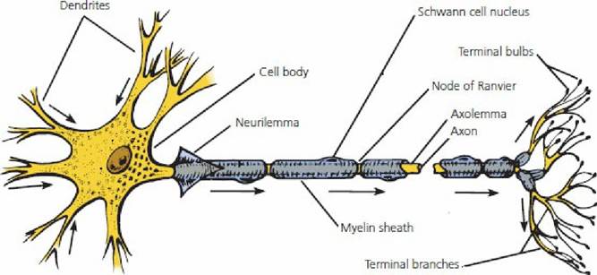
■ FIGURE 4-1 The neuron. Arrows indicate the direction of impulse conduction.
In this myelinated nerve fiber in a peripheral nerve are shown the neurilemma (sheath of Schwann), axolemma (plasma membrane of axon), and nodes of Ranvier. The terminal branches are also referred to as the telodendritic zone. The axon is shown as discontinuous to allow for variable length.The polarity of a neuron refers to the number of poles or processes that stem from its cell body. Mammalian neurons can be categorized as bipolar (one axon and one dendrite extending from the cell body) or multipolar (many branching dendrites and one axon extending from the cell body, see Figure 4-1). Bipolar neurons are found in the retina of the eye and the olfactory region (sense of smell) in the nose (see Chapter 5). Most neurons in the central nervous system (CNS) are multipolar, as shown in Figure 4-1.
The axon (and its myelin covering, if present) is called a nerve fiber. The part of the cell membrane that covers the axon is known as the axolemma. In a myelinated axon, the axolemma is surrounded by a myelin sheath (neurilemma) that is interrupted at regularly spaced intervals by myelin-free gaps, called nodes of Ranvier.
A group of nerve cell bodies within the brain or spinal cord is referred to as a nucleus and a group of nerve cell bodies outside the brain or spinal cord is called a ganglion. A bundle of parallel neuron fibers within the brain or spinal cord is known as either a tract or a fasciculus and a bundle of neuron fibers outside the brain or spinal cord is called a nerve.
Continuity from one neuron to the next is provided by the synapse (Figure 4-2). There is no physical contact of neurons at the synapse. A space exists between the neurons, the synaptic gap, and impulses from one neuron to the next are transmitted by chemical means through this space. This is chemical synaptic transmission, in contrast to electrical synaptic transmission. Because most synaptic transmissions are chemical, our considerations will be limited to chemical transmission. Three notable characteristics of the synapse are: (1) one-way conduction (direction), (2) facilitation (repeated impulses provide for easier subsequent transmission), and (3) greater fatigability than the neuron (allows for repetitive impulses to fade).
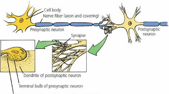
Synaptic gap between terminal bulb and dendrite
■ FIGURE 4-2 The synapse. The enlargements progress in the direction of the arrows.
Glial Cells
Glial cells are the nonneuronal cellular elements of the CNS. They outnumber neurons by about 10fold and make up about half of its volume. The dense packing of neurons and the more numerous glial cells cause nervous tissue to have less interstitial space than other tissues. Glial cells are metabolically quite active.
Glial cells include oligodendrocytes, astrocytes, ependymal cells, and microglia. The most significant role of oligodendrocytes is their involvement in myelin sheath formation in the CNS. A similar function in the peripheral nervous system is performed by Schwann cells. Astrocytes are the most prominent glial cell and their processes abut blood vessels, synaptic structures, and nerve cell bodies and processes. Because of their interposition between blood vessels and neurons, astrocytes not only provide support but also facilitate transport of blood constituents from capillaries to neurons. Also, astrocytes release excitatory neurotransmitter glutamate in response to stimulation. This permits a communication with the neurons by either stimulating or varying its response. Under some circumstances, too much glutamate may be released and lead to excitotoxicity, by which neurons can be killed.
Ependymal cells line the ventricles of the brain and the central canal of the spinal cord. In these locations, the ependymal cells unite with the capillaries to form the choroid plexus, where cerebrospinal fluid is produced.
The microglia have a phagocytic function. They enter the CNS from blood vessels and increase in numbers during inflammatory processes or where neuron injury has occurred.
Myelin Sheaths
Myelin is a white lipid (sphingomyelin) substance that forms a sheath around nerve fibers and serves as an electrical insulator.
It is formed by oligodendrocytes in the CNS and by Schwann cells in the peripheral nervous system (PNS).Nerve fibers within the gray matter of the CNS are not myelinated; their white glistening appearance outside the gray matter, as shown by the white matter and by peripheral nerves, is provided by the myelin that envelops the nerve fibers. Not all nerve fibers outside the gray matter are myelinated, but because of the closeness of unmyelinated fibers to myelinated fibers, they tend to be invaginated (pressed) into the myelin substance. Even when this occurs, however, unmyelinated fibers are uninsulated because they maintain a direct association with extracellular fluid throughout their length.
The Schwann cell cytoplasm (which contains the myelin) is wrapped around a nerve fiber many times, and the nucleus lies within the Schwann cell just beneath the neurolemma external to the myelin sheath (see Figure 4-1). The cytoplasm of the oligodendrocyte is different from that of the Schwann cell because several extensions exist, each of which forms a wrapping around a nerve fiber (Figure 4-3). One cell, therefore, provides a sheath at several locations.
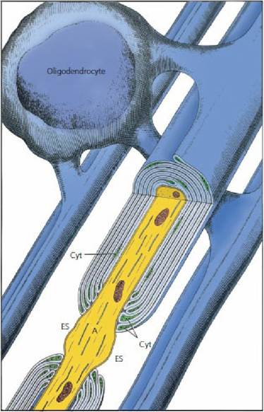
■ FIGURE 4-3 Illustration of an oligodendrocyte providing myelin internodes to three axons. A bisected view of a node, adjacent paranode regions, and part of an internode is shown in the foreground. The axon (A) bulges at the node and is exposed to extracellular space (ES). Loop profiles containing cytoplasm (Cyt) contact the axon at the paranodal regions. (From Eurell JA, Frappier BL. Dellmann’s Textbook of Veterinary Histology. 6th edn. Ames, IA: Blackwell Publishing, 2006.)
Interruptions of the myelin sheath that occur along the length of a fiber are called nodes of Ranvier. These nodes are the junctions of adjacent wrappings, either of the cytoplasmic extensions of oligodendrocytes or of Schwann cells. At these points, the nerve fiber plasma membrane (axolemma) is directly exposed to extracellular fluid.
The exposure is more intimate in the CNS (Figure 4-4). Whereas the sheathed portion of the nerve fiber is insulated, the nodes are uninsulated. Depolarization occurs at the nodes (see the following section) and.the function of the myelin sheaths will become more apparent when nerve conduction is discussed.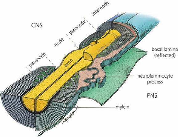
■ FIGURE 4-4 Schematic illustration of nodal and paranodal regions of myelinated fibers from the central nervous system (CNS) (left) and the peripheral nervous system (PNS) (right). In the CNS, myelin is formed by oligodentrocytes and nodes are broadly exposed to the extracellular space. In the PNS, outer cytoplasmic processes of adjacent neurolemmocytes (Schwann cells) overlap to restrict exposure to the extracellular space. (From Eurell JA, Frappier BL. Dellmann’s Textbook of Veterinary Histology. 6th edn. Ames, IA: Blackwell Publishing, 2006.)
■