ORGANIZATION OF THE NERVOUS SYSTEM
1. What are three major subdivisions of the nervous system?
2. What are the three gross divisions of the brain?
3. What are the subdivisions of the brain stem?
4. Is the hypothalamus a subdivision of the cerebrum, cerebellum, or brain stem?
5.
List major characteristics of the cerebral hemispheres, cerebellum, and brain stem.6. What are the five groups of vertebrae in order from their cranial location to their caudal location?
7. What is the vertebral formula for the dog?
8. How are the spinal nerves numbered in relation to the vertebrae?
9. What is the cauda equina?
0. Which root of a spinal nerve does an afferent fiber traverse? An efferent fiber?
11. What are the relative locations of nerve cell bodies and tracts within the spinal cord?
2. What is a spinal cord segment?
3. Are motor neurons located dorsally or ventrally in the gray matter of the spinal cord?
4. What is the general distribution of a spinal nerve?
L5. What is a nerve plexus?
6. How many pairs of cranial nerves are there?
1.7. What is the general distribution of the cranial nerves?
8. Which one of the cranial nerves supplies parasympathetic fibers to visceral structures in the thorax and abdomen?
9. Are all spinal nerves mixed nerves?
0. Are all cranial nerves mixed nerves?
1. What tissues are innervated by autonomic nerves?
!2. Which division of the autonomic nerves is associated with “fight, fright, and flight”?
13. Where are the cells of origin for the sympathetic and parasympathetic neurons?
4. How do autonomic neurons get their structures innervated?
15. Study Table 4-2.
| TABLE 4-1 CRANIAL NERVES | |||
| NO. | NAME | TYPE | DISTRIBUTION |
| I | Olfactory | Sensory | Nasal mucous membrane (sense of smell) |
| II | Optic | Sensory | Retina of eye (sight) |
| III | Oculomotor | Motor | Most muscles of eye |
| Parasympathetic to ciliary muscle and circular muscle of iris | |||
| IV | Trochlear | Motor | Dorsal oblique muscle of eye |
| V | Trigeminal | Mixed | Sensory - to eye and face; motor - to muscles of mastication |
| VI | Abducens | Motor | Retractor and lateral muscles of eye |
| VII | Facial | Mixed | Sensory region of ear and taste to cranial two-thirds of tongue; |
| motor - to muscles of facial expression; parasympathetic - to mandibular and sublingual salivary glands | |||
| VIII | Vestibulocochlear | Sensory | Cochlea (hearing); semicircular canals (equilibrium) |
| IX | Glossopharyngeal | Mixed | Sensory - to pharynx and taste to caudal third of tongue; motor - muscle of pharynx; parasympathetic - to parotid salivary glands |
| X | Vagus | Mixed | Sensory to pharynx and larynx; motor - to muscles of larynx; parasympathetic - to visceral structures in the thorax and abdomen |
| XI | Accessory | Motor | Motor - to muscles of shoulder and neck |
| XII | Hypoglossal | Motor | Motor-to muscles of tongue |
Modified from Frandson RD, Wilke WL, Fails AD.
Anatomy and Physiology of Farm Animals. 7th edn. Ames, IA: Wiley-Blackwell, 2009.
| TABLE 4-2 ACTIONS OF AUTONOMIC STIMULATION | ||
| ORGAN/STRUCTURE | SYMPATHETIC ACTION | PARASYMPATHETIC ACTION |
| Eye | ||
| Muscles of iris | Contraction of radial muscle (dilates pupil) | Contraction of circular muscle (contracts pupil) |
| Heart | ||
| S-A node | Increase in heart rate | Decrease in heart rate |
| A-V node | Increase in conduction velocity | Decrease in conduction velocity |
| Muscle | Increase in force of contraction | Decrease in force of contraction |
| Arterioles | ||
| Skin and mucosa | Constriction | |
| Salivary glands | Constriction | |
| Cerebral | Slight constriction | |
| Skeletal muscle | Dilation | |
| Coronary | Dilation | Slight dilation |
| Pulmonary | Dilation | |
| Abdominal viscera | Constriction | |
| Intestines | ||
| Muscle | Decreased | Increased |
| Secretions | Decreased | Increased |
| Lungs | ||
| Bronchi | Dilation | Constriction |
| Kidney | Afferent arteriole constriction | None |
| Urinary bladder | ||
| Bladder wall | None | Contraction |
| Sphincter | Contraction | Relaxation |
| Penis | Ejaculation | Erection |
| Piloerector muscles | Contraction | None |
| Salivary glands | Mucus secretion | Serous secretion |
All parts of the body are served by the nervous system.
Based on the location of its components, it is subdivided as shown in the following schemes:1. Central nervous system
a. Brain
b. Spinal cord
2. Peripheral nervous system
a. Cranial nerves
b. Spinal nerves
The autonomic nervous system (ANS) is another subdivision of the nervous system and is separate from the above scheme because it has both central and peripheral components with further subdivisions as follows:
1. Autonomic nervous system
a. Sympathetic
b. Parasympathetic
c. Enteric
Central Nervous System
The CNS not only contains components of transmission, but the brain also provides for those functions that we associate with computers, such as memory, a central processing unit for problemsolving, and input-output capability (sensations resulting from sensory input).
The Brain
The gross divisions of the brain are the cerebrum (paired cerebral hemispheres), cerebellum, and brain stem. An organizational scheme (Figure 4-5) shows additional subdivisions. Another scheme (Figure 4-6).is also commonly used; it has different names for the various parts. The relative locations of the various subdivisions to each other according to the first scheme are shown in Figure 4-7.
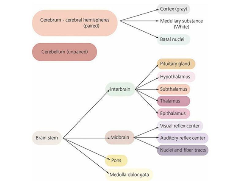
■ FIGURE 4-5 Subdivisions of the brain according to the major divisions, being the cerebrum, cerebellum, and brain stem.
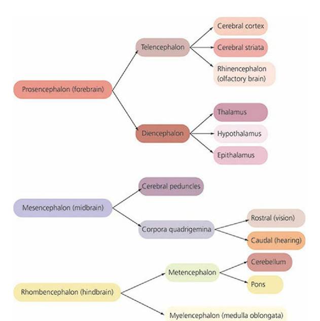
■ FIGURE 4-6 Subdivisions of the brain according to its development from the primary embryonic vesicles, the prosencephalon, mesencephalon, and rhombencephalon.
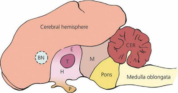
■ FIGURE 4 -7 Relative locations of brain subdivisions to each other. BN, basal nuclei; E, epithalamus; T, thalamus; H, hypothalamus; P, pituitary gland; M, midbrain; CER, cerebellum.
Dotted line for boundaries of basal nuclei represents its location on the midline.Cerebral Hemispheres. The right and left cerebral hemispheres are large structures that make up most of the cerebrum (Figure 4-8). Each hemisphere is composed of a covering of gray matter, the cerebral cortex a central mass of white matter, the medullary substance (made up of nerve fibers); and the basal nuclei (previously known as basal ganglia, but because they are in the CNS, they are now called basal nuclei).
The cerebral cortex has the following characteristics:
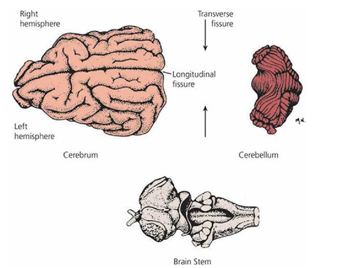
■ FIGURE 4-8 Gross subdivisions of the brain of the dog. (From Beitz AJ, Fletcher TF. The brain. In: Evans HE, ed. Miller’s Anatomy of the Dog. 3rd edn. Philadelphia, PA: WB Saunders Company, 1993.)
1. Acquired late in vertebrate evolution
2. Concerned with those nervous reactions that result in consciousness
3. Regarded as the seat of the highest type of nervous correlation (association)
4. Marked by a high degree of educability (especially in humans)
5. Possesses a motor area:
a. Impulses from these areas in one hemisphere cause muscle movements on the opposite (contralateral) side of the body
b. Size of motor area and number.and complexity of skeletal muscle movements of which an animal is capable are directly related
6. Contains sensory areas, or centers, into which sensory fibers discharge
The sensory areas are: (1) the somesthetic or body sense area, which receives impulses from the skin concerned with touch, warmth, cold, and pain localization; impulses concerned with taste; and impulses from muscles, tendons, and joints; (2) the visual area (sight); (3) the auditory area (hearing); and (4) the olfactory area (smell).
The white matter is composed of myelinated nerve fibers situated beneath the cerebral cortex. These include association fibers, which establish connection between the different parts of the cortex; commissural fibers, which connect the two hemispheres; and projection fibers, which connect the cerebral cortex with other parts of the brain and spinal cord.
The basal nuclei (see Figure 4-7) lie deep within the cerebral hemispheres. They are composed of separate, large pools of neurons organized for the control of complex semivoluntary movements, such as walking and running. In birds the cerebral cortex is poorly developed, but the basal nuclei are highly developed. Because of this contrast, the basal nuclei in birds perform nearly all of the motor functions, even the voluntary movements, in much the same manner as the motor area of the human cortex controls voluntary movement. In the cat, and to a lesser extent in the dog, removal of the cerebral cortex prevents many sophisticated motor functions. Because of the basal nuclei, however, this does not interfere with the ability to walk, eat, fight, and even participate in sexual activity.
Cerebellum The cerebellum (see Figures 4-7 and 4-8) is not concerned with consciousness or,sensation, as is the cerebral cortex. Because of its motor function, the cerebral cortex can start a limb or body part in motion, but once in motion, inertial forces would tend to keep it in motion until opposing forces stopped it. The cerebral cortex is not organized to mobilize the opposing force. The cerebellum, however, can make automatic adjustments to prevent the distortion of inertia and momentum. To accomplish this, the cerebellum receives impulses: (1) from the proprioceptive receptors (located in the internal mass of the body) found in all joints, muscles, and pressure areas (e.g., foot pads); (2) from the equilibrium apparatus of the inner ear; (3) from the visual cortex; and (4) directly from the motor cortex of all motor impulses being sent to muscles (Figure 4-9). Whereas the motor area of a cerebral hemisphere exerts its effect on the opposite (contralateral) side of the body, the effect of one side of the cerebellum is exerted on the same (ipsilateral) side of the body. The cerebellum acts as a “collecting house” for all information regarding the instantaneous physical status of the body.
Cerebellum malfunction is exemplified in feline cerebellar hypoplasia (arrested development) where the mother cat acquires a virus infection while pregnant. Following birth, voluntary muscle movement of the kittens overreaches the intended goal (hypermetria); there is failure of muscle, coordination (ataxia) and involuntary trembling or quivering (tremor). A very underdeveloped cerebellum is revealed when the kittens are necropsied.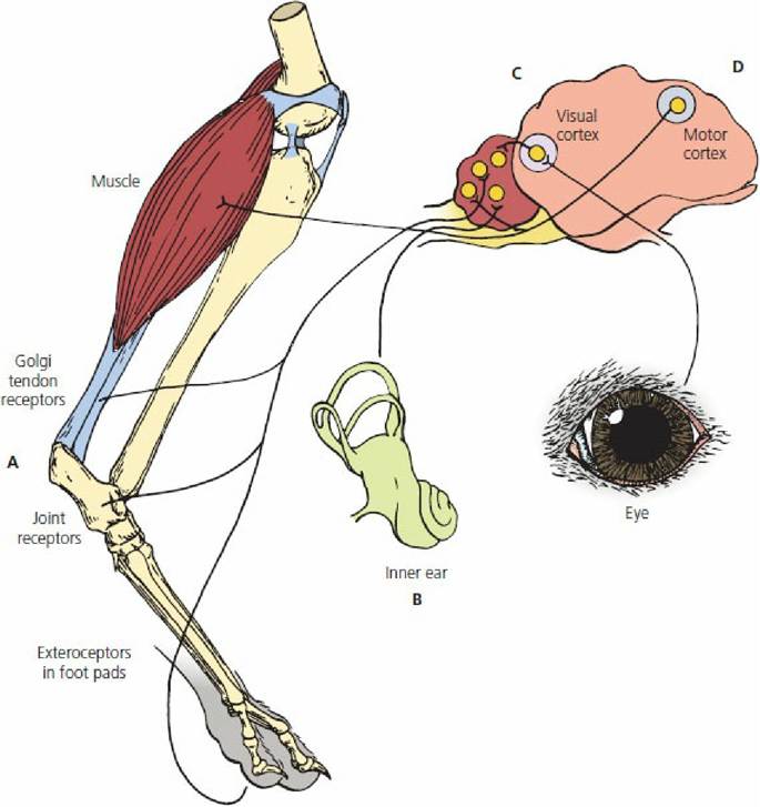
■ FIGURE 4-9 Sources of input to the cerebellum of the dog. A. Exteroceptors in foot pads (pressure) and proprioceptors in joints, muscles, and tendons (tension). B. Vestibular apparatus (equilibrium) of the inner ear. C. Visual cortex of the cerebrum. D. Cerebral motor cortex (simultaneous impulse to muscle).
Brain Stem
The brain stem is composed of the interbrain rostrally followed caudally (in order) by the midbrain, pons, and medulla oblongata (see Figures 4-7 and 4-Figure 4-8). The cerebral hemispheres and cerebellum arise from the brain stem. In addition to the many fiber tracts that ascend and descend between the spinal cord and the cerebrum and cerebellum, the brain stem is the origin of all the cranial nerves except for the optic, olfactory, and acoustic nerves (special senses). The cells of origin for the latter lie outside of the skull.
From below upward, the interbrain is composed of the hypothalamus, thalamus, and epithalamus (see Figure 4-7). The hypothalamus contains the hypophysis or pituitary gland, which is an endocrine organ. Associated with the hypothalamus is a complex sensing and neurosecretory function. Also, the hypothalamus assumes a major role in the integration of functions carried out by the autonomic nervous system. For these functions, the anterior and middle portions contain parasympathetic components and the posterior portion contains sympathetic components. The thalamus contains many nuclei and is truly a relay center. Impulses from all areas of the body are transmitted to the thalamus for transfer to the cerebral cortex. Other nuclei in the thalamus are associated with the relay of impulses within the brain. The epithalamus contains an olfactory (smell) correlation center and the pineal gland. The latter is a neurosecretory organ that regulates gonadal hormones and certain daily rhythms.
The midbrain (see Figure 4-7) contains the auditory and visual reflex centers, the nuclei of two cranial nerves, and several descending tracts.
The medulla oblongata and pons (see Figure 4-7) contain many ascending and descending pathways, the sensory and motor nuclei for all of the cranial nerves originating in the brain stem (except the two located in the midbrain), and a large part of the central mechanism of the postural reflexes (e.g., hopping, righting, placing). There are also several reflex centers associated with the regulation of important visceral functions such as heart rate, blood vessel muscle tone (vasomotor tone), respiration, and motor and secretory activities of the digestive tract.
Spinal Cord
The spinal cord is the caudal continuation of the medulla oblongata. Segmentation (association with the vertebral segments) is noticeable, with each segment giving rise to a pair of spinal nerves. The spinal cord receives sensory afferent (inflowing) fibers by way of the dorsal roots of the spinal nerves and gives off efferent (outflowing) motor fibers to the ventral roots of the spinal nerves (Figure 4- 1O).
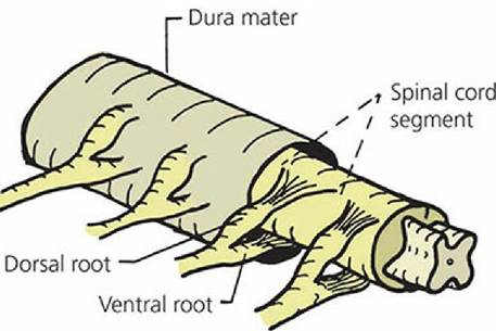
■ FIGURE 4-10 Structure of the spinal cord of the dog, showing a spinal cord segment. (Redrawn from Breazile JE. Textbook of Veterinary Physiology. Philadelphia, PA: Lea & Febiger, 1971.)
The centrally located gray matter (which resembles a capital H and is sometimes called the gray H) consists primarily of nerve cell bodies and their processes. The peripherally arranged white matter, which has a white appearance because of its myelin sheaths, is composed of many distinct tracts (Figure 4-11). A tract is a bundle of nerve fibers having a common origin, termination, and function, and connects the brain stem and higher centers with the spinal nerves. Different sensory and motor tracts are segregated in the cord. Proprioceptive (referring to the sensing of position of limbs or other body parts without the use of vision) impulses from muscles, tendons, and joints have well- defined ascending tracts, as do sensory impulses for pain, temperature, and touch. Similarly, impulses associated with certain motor functions descend in definite tracts. Many of the tracts are named according to the structures they connect. For example, the ventral spinocerebellar tract carries impulses from the spinal cord to the cerebellum. The lateral spinothalamic tract carries impulses from the spinal cord to the thalamus. The cells of origin for sensory impulses to the brain or to other parts of the spinal cord are located in the dorsal horns of the gray matter, and the cells of origin of motor impulses to the spinal nerves are located in the ventral horns of the gray matter. The cells of origin of the autonomic motor impulses arising from the spinal cord are the lateral masses of the ventral horns (intermediate location) of the gray matter (see Figure 4-11).
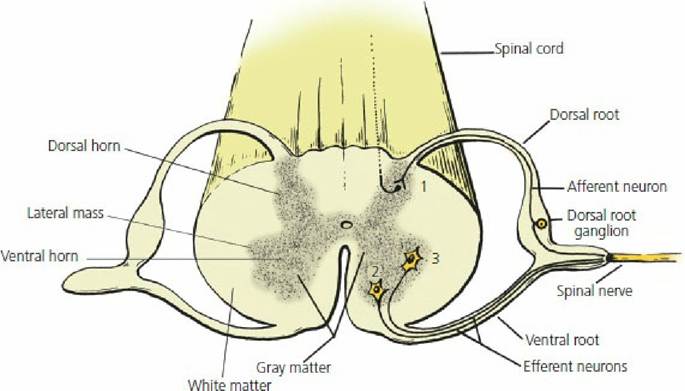
■ FIGURE 4-11 Transverse section of the spinal cord of the dog. Located within the gray matter are: (1) nerve cell bodies for sensory neurons in dorsal horns; (2) somatic motor neurons in ventral horns; and (3) autonomic motor neurons in lateral masses of ventral horns.
As the spinal cord descends and proceeds caudally, its cross-sectional area decreases. Finally, at the caudal extremity, the tracts terminate and the spinal nerves fan outward and backward, giving the appearance of a broom or horse’s tail. Accordingly, the terminal part of the spinal cord, meninges, and nerves is called the cauda equina (Figure 4-12).
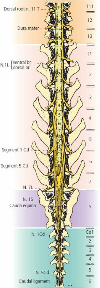
■ FIGURE 4-12 Caudal extremity of the spinal cord showing the cauda equina. Vertebrae numbers are designated on the right and the spinal cord segments are identified within the drawing of the cord. T = thoracic, L = lumbar, S = sacral, Co = coccygeal. (From Fletcher TF, Kitchell RL. Anatomical studies on the spinal cord segments of the dog. Am J Vet Res. 1966; 27: 1762.)
Peripheral Nervous System
The peripheral nervous system consists of the spinal nerves and the cranial nerves.
Spinal Nerves
Spinal nerves, as well as cranial nerves, are referred to as somatic nerves because of their association with voluntary control of muscles. Autonomic nerves are referred to as visceral nerves because they are involved with involuntary functions such as control of smooth muscle, cardiac muscle, and glands. The spinal nerves are those that arise from the spinal cord and emerge from the vertebrae. In the dog, for example, there are 7 cervical, 13 thoracic, 7 lumbar, 3 sacral, and an average of 20 caudal vertebrae. With the exception of the cervical and caudal nerves, there is a pair of spinal nerves (one right and one left) that emerges behind the vertebrae of the same serial number and name. In this plan the first pair of thoracic nerves emerges through the intervertebral foramina located between the Ti and T2 vertebrae, and the last pair of thoracic nerves emerges through the intervertebral foramina between the T13 and L1 vertebrae (Figure 4-13B and C). There are the same number of pairs of thoracic, lumbar, and sacral nerves as there are similar vertebrae. Instead of seven pairs of cervical nerves (corresponding with seven cervical vertebrae), however, there are eight pairs. The first pair of cervical nerves emerges through the foramina in the C1 vertebra and the second pair emerges between the C1 and C2 vertebrae.(Figure 4-13A). Usually there are only six or seven pairs of caudal nerves.
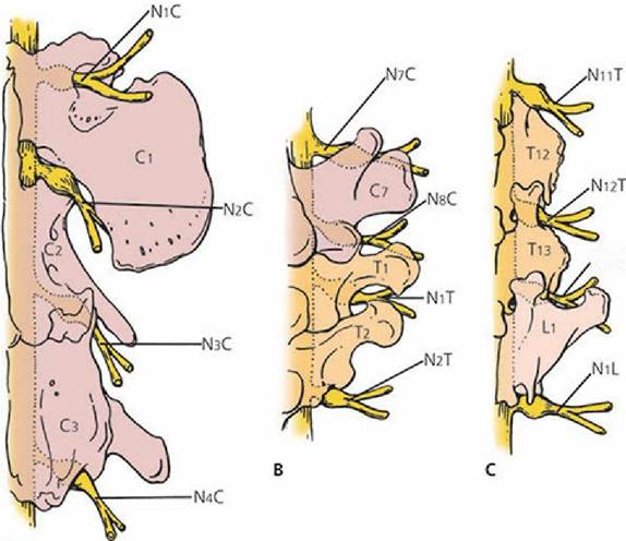
A
■ FIGURE 4-13 A schematic representation of the association of spinal nerves with vertebrae in the dog. Only the right half of the spinal cord, vertebrae, and spinal nerve pair is shown. A. C1 to C3 vertebrae. B. C7, T1, and T2 vertebrae. C. T12, T13, and L1 vertebrae. Although not consistently shown, the dorsal root ganglions are medial to the emergence of spinal nerves through intervertebral foramina.
A spinal nerve is composed of a dorsal and ventral root and its branches. The dorsal root enters the dorsal portion of the spinal cord. It carries afferent (sensory) impulses from the periphery toward the spinal cord (see Figure 4-11). The nerve cell bodies of the neurons composing the dorsal root are located in the dorsal root ganglion (DRG). This is visible as an enlarged part of the dorsal root close to the point where the dorsal and ventral roots join to form the spinal nerve proper.
The ventral root emerges from the ventral horn of the spinal cord. It carries efferent (motor) impulses from the spinal cord to striated muscle fibers (see Figure 4-11). Near the intervertebral foramen, the dorsal root joins with the ventral root to form the main part of the spinal nerve. The spinal nerve proper is classified as a mixed nerve because it contains both sensory and motor fibers. After the spinal nerve emerges from the intervertebral foramen, it divides into a dorsal branch and a ventral branch; these supply innervation to structures dorsal and ventral to the transverse processes of the vertebrae, respectively (Figure 4-14). The spinal nerves generally supply sensory and motor fibers to the region of the body in the area where they emerge from the spinal cord, but this is not the case for the appendages. They are innervated by the ventral branches of several spinal nerves and, near the limb they supply, the nerves join together in braid-like arrangements known as plexuses. Each forelimb is supplied by nerves that arise from the brachial plexus (Figure 4-15) and each hindlimb is supplied by nerves that arise from the limbosacral plexus.
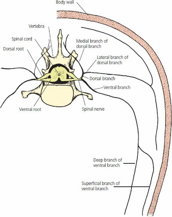
■ FIGURE 4-14 A spinal nerve and its location relative to its branches, roots, spinal cord, and vertebra.
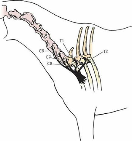
■ FIGURE 4-15 Brachial plexus of the horse. It is formed by the contributions of the last three cervical and first two thoracic spinal nerves to supply the forelimbs. C, cervical; T, thoracic. The corresponding numbers refer to their respective spinal nerve.
Cranial Nerves
There are 12 pairs of cranial nerves, with a right and a left nerve making up each pair. The cranial nerves usually supply innervation to structures in the head and neck. The vagus nerve is an exception. In addition to its sensory and motor supply to the pharynx and larynx, it also supplies parasympathetic fibers to visceral structures in the thorax and abdomen (Figure 4-16). These nerves have no dorsal or ventral roots and emerge through foramina in the skull. Some cranial nerves are strictly sensory (afferent), some are strictly motor (efferent), and some are mixed (both sensory and motor). The cranial nerves are listed by number, name, type, and distribution in Table 4-1.
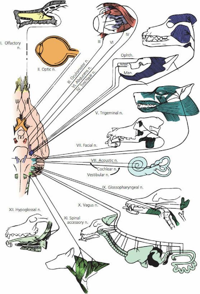
■ FIGURE 4-16 Origin and major distribution of cranial nerves in the dog. N, nerve; OPHTH, ophthalmic nerve; MAX, maxillary nerve; MAN, mandibular nerve. Acoustic nerve (VIII) now called vestibulocochlear nerve and spinal accessory nerve (XI) now called accessory nerve. (From Hoerlein BF, Oliver JE, Mayhew JG. Neurologic examination and the diagnostic plan. In: Oliver JE, Mayhew IG, eds. Veterinary Neurology. Philadelphia, PA: WB Saunders Company, 1987.)
Autonomic Nervous System The ANS, also known as the involuntary, vegetative, or visceral nervous system, is essential for maintaining normal organ function (homeostasis), adapting to,environmental change (e.g., body temperature), and responding to stresses (e.g., excitement). These responses are accomplished by either simple or complex reflexes and with little, if any, conscious perception. The ANS has sympathetic, parasympathetic, and enteric subdivisions: (1) the sympathetic nervous system (SNS) is associated with the body’s response to stress; (2) the parasympathetic nervous system (PSNS) is associated with homeostatic functions in the absence of stress; and (3) the enteric nervous system (ENS) is associated with regulation of the gastrointestinal (digestive) system. The ENS functions mostly by itself but can be modulated by both the SNS and PSNS. The ENS will be described in more detail in Chapter 12. These subdivisions have mostly efferent activities, but not to be overlooked are the many sources of afferent information needed for its reflex functioning.
ANS innervation extends to smooth muscle, cardiac muscle, and glands. Most organs receive both sympathetic and parasympathetic innervation. Exceptions are sweat glands, most blood vessels, the uterus, and piloerector muscles of the skin, all of which receive only sympathetic innervation. The effect of sympathetic stimulation is generally opposite to that of parasympathetic stimulation. Accordingly, they can be considered as antagonistic to each other. To visualize their respective functions in regard to an organ, the so-called “fight, fright, or flight” concept can be considered. Those actions that would be considered favorable in a fighting, frightening, or retreating situation can be attributed to sympathetic activity, and those actions associated with restful or tranquil situations (eat-or-sleep concept) can be attributed to the parasympathetic activity. A comparison of their respective actions for various organs is presented in Table 4-2.
Central Components
The central component resides in the central processing that results from data received (e.g., blood pressure, organ distention) from afferent (inflowing) neurons. The central processing also relies on a number of blood signals such as temperature, pH, glucose concentration, and others. The central processing for the reflex integration and modulation of body conditions by the ANS occurs in spinal cord sites and a number of brain locations. The latter is best represented by the hypothalamus. After central processing, reflex adjustments are made by the peripheral (efferent) components of the ANS.
Peripheral Components
The cells of origin for the sympathetic nerves are located in the lateral masses of the ventral horns of the thoracic and lumbar segments of the spinal cord, and the cells of origin of the parasympathetic nerves are located in the brain stem and sacral segments of the spinal cord; hence the term for their origin is noted as craniosacral for the parasympathetics as opposed to thoracolumbar for the sympathetics (Figure 4-17). For both sympathetic and parasympathetic activity, two neurons (in series) are associated with the transmission of impulses from the cells of origin in the spinal cord or brain to the effector organ (glands or muscle). The cells of origin for the second neuron are located in ganglia. The first neuron is called preganglionic and the second neuron is called postganglionic.
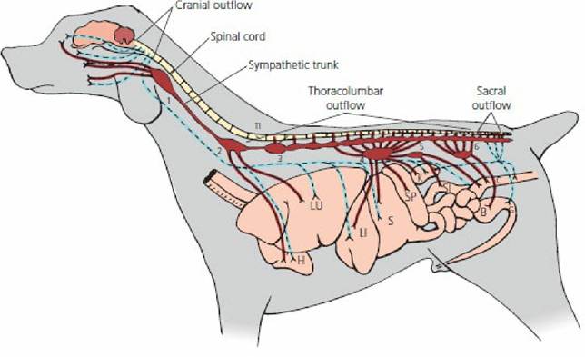
■ FIGURE 4-17 Diagrammatic representation of the efferent autonomic nervous system of the dog. Only one chain of the bilateral sympathetic trunk is shown. The lines showing sympathetic outflow (thoracolumbar) are red; lines for parasympathetic outflow (craniosacral) are blue. Numbers indicate sympathetic ganglia: (1) cranial cervical; (2) middle cervical; (3) cervicothoracic; (4) celiac; (5) cranial mesenteric; (6) caudal mesenteric. LU, lung; H, heart; LI, liver; S, stomach; SI, small intestine; SP, spleen; K, kidney; C, colon; B, urinary bladder; G, genitalia; T1, 1st thoracic vertebra.
Sympathetic Efferent Distribution
The preganglionic neuron for a sympathetic nerve traverses the ventral root of a thoracic or lumbar spinal nerve, enters the spinal nerve proper, and soon branches from it to enter a vertebral ganglion of the sympathetic trunk, a bilateral chain of ganglia ventral to the vertebrae (see Figure 4-17) with a ganglion on each side of each vertebrae. It either synapses in a ganglion of the same vertebral segment or it can continue over a considerable distance to another vertebral ganglion, where it synapses. The synapse might not occur in a vertebral ganglion at all, however, but might continue to some paired ganglia that are ventral to the sympathetic trunk; these are called prevertebral ganglia. The prevertebral ganglia are fewer in number and include the cranial cervical ganglion (with distribution to smooth muscle and glands of the head), middle cervical ganglion (heart and lungs), cervicothoracic ganglion (arteries in neck and thorax), celiac ganglion (stomach, liver, pancreas, kidney, adrenal), cranial mesenteric ganglion (small intestine and upper colon), and caudal mesenteric ganglion (lower colon and neck of the bladder). The postganglionic neuron leaves the vertebral or prevertebral ganglion and proceeds to the effector organ, usually by way of a blood vessel to that organ. It can also leave the vertebral ganglion, reenter a spinal nerve, and be distributed by the branchings of the spinal nerve.
Parasympathetic Efferent Distribution
The preganglionic neurons of the parasympathetic division are distributed to ganglia near the effector organs before they synapse with the postganglionic neuron. Accordingly, the preganglionic fibers are relatively longer and the postganglionic fibers are relatively shorter as compared with the preganglionic and postganglionic fibers of the sympathetic division. Most of the parasympathetic ganglia are microscopic and are an intimate component of the tissue they innervate. The parasympathetic preganglionic fibers that arise from nerve cell bodies in the brain are distributed to their respective organs in common with one of four cranial nerves (III, VII, IX, or X). The first three supply regions of the head, and the last, cranial nerve X (the vagus nerve), supplies the heart and lungs in the thorax and nearly all of the abdominal viscera (see Figure 4-17). The vagus nerve has sometimes been called the vagabond nerve because of its extensive wanderings. The parasympathetic preganglionic fibers that arise from nerve cell bodies in the sacral portion of the spinal cord supply the last part of the digestive tract and most of the urogenital system (see Figure 4-17). These fibers emerge from the ventral branches of their respective segments and are distributed to the ganglia near the effector organs supplied by the pelvic nerve.
Autonomic Reflexes
The autonomic function is based on reflex activity, and these reflexes control such functions as blood pressure, heart rate, and the activity of the digestive and urogenital systems (see Table 4-2). Autonomic reflexes involve afferent transmission of sensory information from effector organs to the CNS, information processing, and return of a motor response to the effector organs. Autonomic afferents are not designated as sympathetic or parasympathetic (i.e., they transmit information regardless of which division of the ANS) and most travel to the CNS via SNS and PSNS nerves. Their cell bodies are in DRG and cranial nuclei. Some afferents (e.g., blood vessels in skeletal muscle) travel in spinal and cranial nerves. Most of the autonomic functions do not reach the conscious level. However, some afferent information carried by autonomic sensory neurons do reach conscious levels. This may be normal or pathologic. Normal would include feelings of fullness of the bladder or rectum and pathologic might include gallbladder pain or angina pectoris as experienced by humans.
N eurotransmitters
A nerve impulse causes an effect at a synapse or at the structure being innervated. Axons terminate by branching; the branches terminate with a structure known as a presynaptic terminal bulb at the synapse and with other similar, modified structures at the organs innervated (see Figure 4-2). These terminations have vesicles containing chemical substances that are liberated when the impulse arrives. The chemical substance then diffuses to the membrane of the postsynaptic neuron or structure and influences the permeability of the membrane for sodium ions.
Peripheral Neurotransmitters
The neurotransmitters of the somatic peripheral nervous system are excitatory in nature - that is, they increase the permeability of the affected membrane for sodium ions. This substance is acetylcholine (ACh) for the somatic spinal and cranial nerves. ACh is also the preganglionic and postganglionic terminal neurotransmitter for the parasympathetic division of the autonomic nervous system (Figure 4-18). This division of the autonomic nervous system is, therefore, sometimes referred to as the cholinergic system. The preganglionic terminal neurotransmitter of the sympathetic division is also ACh, but the postganglionic terminal secretion is usually norepinephrine. Another name for norepinephrine is noradrenaline, so the sympathetic division is often referred to as the adrenergic system.
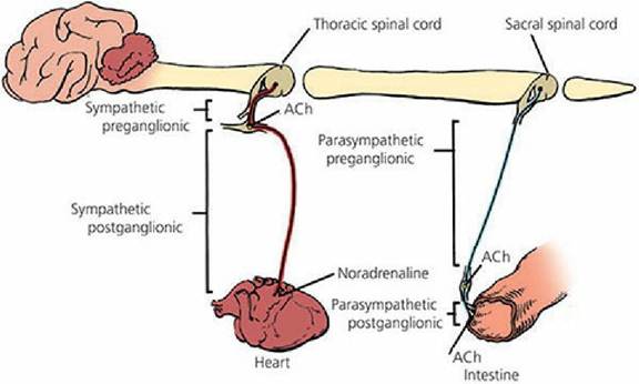
■ FIGURE 4-18 The neurotransmitters acetylcholine (ACh) and norepinephrine (noradrenaline) associated with the autonomic nervous system of mammals.
Central Neurotransmitters
In the central nervous system there are not only excitatory but also inhibitory transmitters. In addition to ACh and norepinephrine, which are present in peripheral neurons, other excitatory transmitters are found in the central nervous system. At least two inhibitory transmitters are recognized within the brain and spinal cord, gamma-aminobutyric acid (GABA) and glycine, which is a simple amino acid. A decrease in the permeability of the affected membrane for sodium ions is one mechanism of inhibition.
■