SUPERFICIAL NERVES
The distribution of the cutaneous nerves follows the general pattern (see Figures 8-68 and 8-69). The dorsal branch (Figure 11-6, A/7-B/7) of the facial nerve runs across the dorsal half of the masseter; the ventral branch takes the more protected course along the ventral edge.
They are joined together by communicating branches at the rostral border of the muscle. The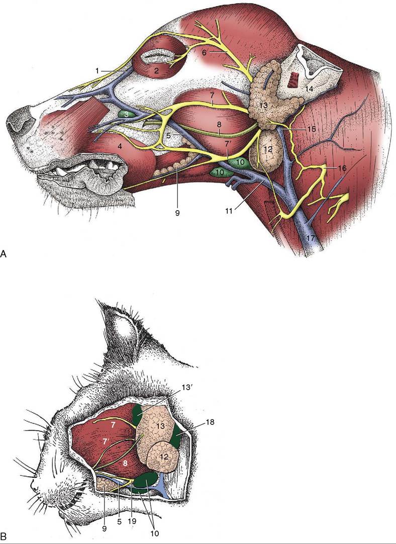
Figure 11-6 Superficial dissection of the canine (A) and feline (B) heads. 1, Angularis oculi vein; 2, orbicularis oculi; 3, facial lymph node; 4, orbicularis oris; 5, facial vein; 6, auriculopalpebral nerve; 7,7’, dorsal and ventral buccal branches of facial nerve; 8, parotid duct; 9, buccal salivary glands; 10, mandibular lymph nodes; 11, linguofacial vein; 12, mandibular gland; 13, parotid gland; 13', parotid lymph node; 14, base of ear; 15, maxillary vein; 16, second cervical nerve; 17, external jugular vein; 18, retropharyngeal lymph node; 19, facial nerve, ventral branch.
auriculopalpebral branch of the facial nerve (Figure 11—6/6) passes across the zygomatic arch, where it can be blocked to eliminate blinking (m. orbicularis oculi) during examination of the eye.