Sympathetic/ thoracolumbar division
Key points
■ The sympathetic nervous system originates from the lateral/intermediate horn of the thoracolumbar spinal cord.
■ Peripheral ganglia are located in the paired, paravertebral sympathetic trunks, or the median, prevertebral ganglia in the dorsal aspect of the thoracic and abdominal cavities.
■ Postsynaptic neurons travel via spinal nerves, or specific named nerves, to their target organ.
■ Visceral structures in the head are supplied by postsynaptic neurons originating in the cranial cervical ganglion.
■ The thoracic viscera are innervated by neurons primarily originating in the cervicothoracic and middle cervical ganglion.
■ The abdominal and pelvic regions are supplied by branches from the thoracic and abdominal sympathetic trunks, and prevertebral ganglia located around the aorta.
Presynaptic fibres originate in the intermediate (lateral) horn of the thoracolumbar spinal cord. Fibres may leave in the ventral root from their spinal cord segment of origin, or they may pass cranially, or caudally, a number of segments within the spinal cord before exiting it. The fibres exit the spinal cord along with the somatic motor neurons using the ventral roots of C8-L4/5 (up to L7) spinal nerves. The ventral roots fuse with the dorsal roots to form proper spinal nerve at the level of the intervertebral foramen (see Fig. 1.1). Lateral to the foramen, the proper spinal nerve splits into epaxial, hypaxial and ventral branches. The ventral branch forms the ramus communicans (ramus - L = branch). The ramus communicans conveys sympathetic efferent and visceral afferent fibres, between the spinal cord and the bilateral, sympathetic trunks that run ventrolaterally on both sides of the vertebral column (Fig. 12.3). The ramus communicans conveys both presynaptic (myelinated) sympathetic efferent fibres that are travelling to the trunk, and postsynaptic (unmyelinated) fibres that return from the trunk to rejoin the spinal nerves.
This gives rise to the names of white and grey ramus communicans, respectively. These paravertebral ganglia and trunks are prominent in the thoracic region and extend into the lumbar region (see Fig. 12.4). In the caudal lumbar area, the trunks may fuse and continue caudally, ventral to the sacral and caudal vertebrae. In the abdominal region, nerves leave the paravertebral sympathetic trunks and connect to prevertebral ganglia located ventral to the vertebral column near the large abdominal arteries. Cranially, the sympathetic fibres continue into the cervical region, in conjunction with the vagus nerve forming the vagosympathetic trunk; this is located in the carotid sheath.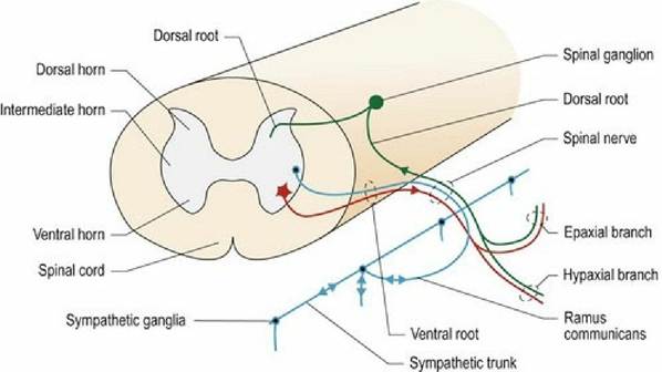
Fig. 12.3 Sympathetic efferent and visceral afferent fibres connecting between the thoracolumbar spinal cord and the sympathetic trunk.
A ganglion occurs where each ramus communicans joins the sympathetic trunk. The ganglia are numbered based on the spinal nerve that supplies them. Fibres may pass through several ganglia before synapsing. Thus the sympathetic trunk comprises both pre- and postsynaptic fibres; these are myelinated and unmyelinated, respectively. From the paravertebral ganglia, there are a number of routes that the fibres may take.
(a) Return via the ramus communicans to join spinal segmental nerves to be distributed to somatic targets such as blood vessels or skin (glands).
(b) Travel further ventrally in specific nerves to innervate viscera. For example, fibres from the cervicothoracic ganglion go to the heart via cardiac nerves.
(c) Travel cranially or caudally to other ganglia and then peripherally, via other spinal nerves.
(d) Travel along arteries to supply a distant part of the body. For example, fibres from the cranial cervical ganglion travel rostrally associated with arteries of the head region. Note that sympathetic fibres also supply the walls of these blood vessels and thereby have an important role in circulatory regulation.
Supply to the head and thorax
On each side, ganglia in the caudal cervical and cranial thoracic region have fused to form the cervicothoracic (stellate) ganglion and the more ventrally located, middle cervical ganglion. Sympathetic fibres from T1-T4 (T5) supply the cervicothoracic ganglion, located in the dorsal thorax at the level of the first rib. Some fibres synapse here, but fibres supplying the neck and head pass through it without synapsing. This is the largest autonomic ganglion in the body. Cranial to the cervicothoracic ganglion the sympathetic trunk splits forming the ansa subclavia (ansa - L = handle or loop). The ansa subclavia passes ventrally around the subclavian artery and connects with the middle cervical ganglion. Fibres run cranially from each middle cervical ganglion in the vagosympathetic trunk, to supply the neck and head. They synapse in the cranial cervical ganglion near the tympanic bulla. In horses, the ganglion is located in the mucous membrane of the guttural pouch (medial compartment), thus it is susceptible to bystander injury in guttural pouch diseases. In dogs, the postsynaptic neurons pass through the tympano-occipital fissure between the tympanic bulla and the petrosal bone, along with the internal carotid artery. They join the ophthalmic branch of the trigeminal nerve, passing through the orbital fissure to be distributed to the eye. Other axons leave the cranial cervical ganglion and are distributed to the blood vessels and glands of the head, while some fibres may travel with branches of cranial nerves IXXI to supply the larynx and pharynx.
The sympathetic supply to the head innervates smooth muscle (vascular, ocular and orbital, erector pilae) and glands (sweat, salivary, nasal).
Sympathetic innervation of the thoracic viscera is derived from the cervicothoracic and middle cervical ganglia. Fibres join with those of the vagus nerve for distribution to thoracic organs.
Supply to the abdomen
Arising from the thoracic and abdominal sympathetic trunk, thoracic and lumbar splanchnic nerves supply pre- and postsynaptic fibres to the abdominal and pelvic regions. These nerves also carry afferent fibres from the viscera. The specific anatomy varies between individual animals. The splanchnic fibres travel to prevertebral ganglia located ventral to the aorta and its branches. These ganglia include the obvious celiac, cranial and caudal mesenteric ganglia, as well as the less obvious adrenal, phrenico-abdominal, renal and gonadal ganglia. Some ganglia are paired and some have contributions from the dorsal branch of the vagus nerve. Hypogastric nerves arising from the caudal mesenteric ganglia are postsynaptic; they supply the pelvic viscera with sympathetic innervation.