Terminology
Certain terms (Fig. 4-3) routinely used in reference to bones, particularly long bones, include the following.
Compact (dense or cortical) bone is the hard layer that constitutes the exterior of most bones and forms almost the entire shaft of long bones.
Cancellous (spongy) bone is composed of spicules arranged to form a porous network. The spaces are usually filled with marrow.
The medullary cavity (marrow cavity) is the space surrounded by the cortex of a long bone. in young animals it is filled with red marrow (hematopoietic tissue), which gradually is replaced by yellow marrow (fat) as the animal ages.
Epiphysis refers to either end of a long bone. The end closest to the body is the proximal
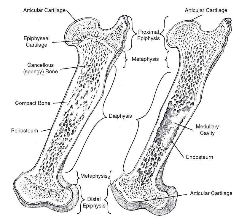
Figure 4-3. Longitudinal section of the equine femur. Left) Immature (growth plates open). Right) Mature (growth plates fused).
epiphysis, and the end farthest from the body is the distal epiphysis.
The diaphysis is the cylindrical shaft of a long bone between the two epiphyses.
The metaphysis of a mature bone is the flared area adjacent to the epiphysis.
Epiphyseal cartilage or disk (physis) is a layer of hyaline cartilage within the metaphysis of an immature bone that separates the diaphysis from the epiphysis. This is the only area in which a bone can lengthen.
Articular cartilage is a thin layer of hyaline cartilage that covers the articular (joint) surface of a bone.
Periosteum is a fibrous membrane that covers the surface of a bone except where articular cartilage is located. Osteoblasts (bone-producing cells) of the periosteum are responsible for increases in the diameter of bones, and activity of periosteal cells is important in the healing of fractures. The periosteum is vascular and well-innervated.
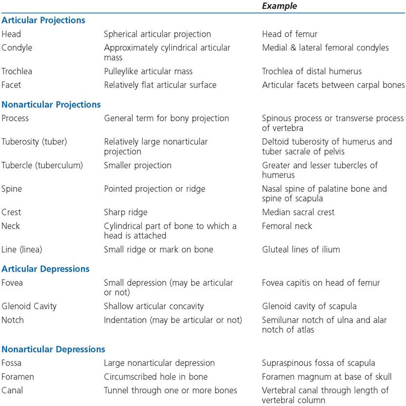
Table 4-1. Bony Features
Endosteum is a fibrous membrane that lines the marrow cavity and osteonal canals (osteons) of a bone. Erosion of existing bone by osteoclasts (bone-destroying cells) in the endosteum determines the size of the marrow cavity and the thickness of the diaphyseal cortex. Both periosteum and endosteum contain osteoblasts and osteoclasts (see Chapter 5).
Many of the projections from and depressions in bones have general names that depend to some extent on their size and function. Both projections and depressions may be articular or nonarticular. if they are articular, they form an integral part of a joint and are covered with articular cartilage. Nonarticular projections and depressions exist outside of joints. Many of them provide areas for attachment of muscle tendons or of ligaments. Table 4-1 lists some common bony features.