TESTES
Spermatozoa are produced by the testes. They have a similar structure, even if they differ slightly in size, shape, and position across species. The majority of each testicle is occupied by the convoluted seminiferous tubules.
Within them, spermatozoa are created. The tunica albuginea is a capsule made of connective tissue that encloses the testicle. The tunica albuginea’s connective tissue extensions, referred to as septa or trabeculae, support the seminiferous tubules inside the testis. The link between the seminiferous tubules and their connective tissue support (interstitial tissue) is revealed in a cross-section of the testicles (Figure 20.1).20.2.1 Sertoli Cell (Sustentacular Cell) and
the Leydig cell (Interstitial Cell)
To help developing spermatozoa, the Sertoli cell acts as a “nurse.” Sertoli cell processes encircle spermatids and spermatocytes, establishing close contact with them during the entire spermatozoa generation process; for this reason, they are referred to as sustentacular, or supporting, cells. The Sertoli cells extend towards the center of the seminiferous tubules from their base at their periphery. The blood-testis barrier is created by the basal junction, also known as the tight junction, forming with nearby Sertoli cells. This barrier allows control over the tubule’s environment and prevents spermatozoa from entering the interstitium.
The seminiferous tubules are divided into two compartments by the Sertoli cells: the adluminal compartment, which is the space between Sertoli cells and which communicates centrally with the tubule lumen, and the basal compartment, which provides space to germinal epithelial cells and interacts with interstitial fluid. A replacement cell and another cell that needs to pass through the Sertoli cell junction in order to access the adluminal compartment are produced when a germinal epithelial cell (spermatogonium) divides in the basal compartment.
Here, more divisions take place, and the final spermatozoa are produced. A fluid that is secreted into the adluminal compartment by the Sertoli cells is advantageous to the growing spermatozoa. The Leydig cells are found in the connective tissue surrounding the seminiferous tubules and are responsible for testosterone production (Figure 20.2).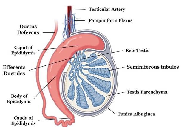
FIGURE 20.1 Cross-section of the testicles
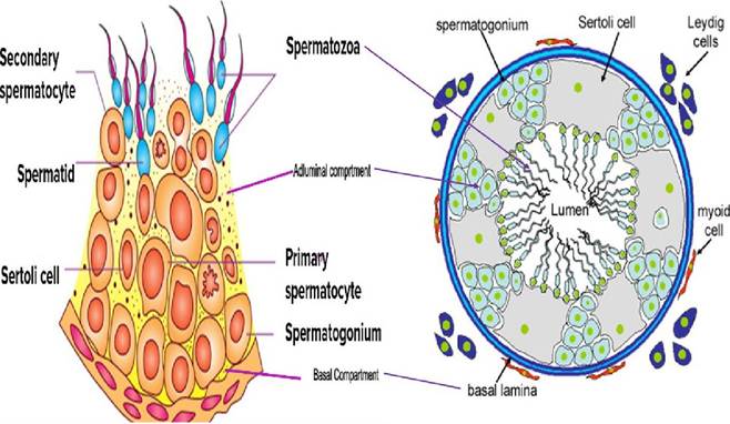
FIGURE 20.2 Illustrative diagram showing sertoli cells and Leydig cells
20.2.2 Epididymis
The testis’s collecting and storing tubule is called the epididymis. The head of the epididymis, where blood vessels and nerves enter at the pole of the testis, is where it starts. The body of the epididymis, which eventually ends and turns upward to become the tail, extends from the head along one side of the testis (Figure 20.1).Sperm and adlu- minal fluid from the rete testis are delivered to the head of the epididymis via efferent ducts. Rete testis is the intra- testicular network of straight tubules that receives content from the convoluted seminiferous tubules. Fluid from the adluminal gaps enters the lumen of the seminiferous tubules and transports spermatozoa to the epididymis. The spermatozoa mature and become motile while being stored in the epididymis.
A large portion of the seminiferous tubular fluid is reabsorbed in the epididymis head.
20.2.3 Ductus Deferens
The ductus deferens, also known as the vas deferens, is the duct system that extends from the pelvic urethra to the tail of the epididymis. Within the visceral layer of the vaginal tunic, the ductus deferens, testicular artery, vein, nerve, and lymphatic vessels are encapsulated as they leave the testis and move towards the abdomen. The spermatic cord is the name given to this group of components. The visceral layer of the vaginal tunic also envelops the testis and epididymis.
It originates from the embryonic abdominal peritoneum that was formed when the testes passed through the inguinal canal and reached the scrotum. The inguinal canal, which runs from the interior to the exterior inguinal rings, is an oblique passageway that connects the abdominal cavity to the outside of the body. The tendinous attachments of the two flat abdominal muscles to the pelvis include gaps called inguinal rings.The ductus deferens splits from the spermatic cord to reach the pelvic urethra after the spermatic cord travels through the inguinal rings. The ampulla of the ductus deferens, which is lacking in the dog and boar, is an expanded, glandular region at the end of the ductus deferens that varies in size among species.
20.2.4 Scrotum
The testes are located in a cutaneous bag called the scrotum. The subcutaneous layer of smooth muscle fibers in the scrotum, known as the tunica dartos, contracts during the colder months to draw the testes closer to the abdominal wall. The parietal layer of the vaginal tunic, which extends the parietal peritoneum into the scrotum, lines the inside of the scrotum. During embryonic development, the testes are intra-abdominal but outside the peritoneum. They have not yet entered the scrotum, but each has a fibrous connection to the scrotum known as the gubernaculum testis. As development and growth progress, the gubernaculum testis “pulls” the testes through the inguinal canal into the scrotum, which creates a double-walled tube of peritoneum. The testis, epididymis, ductus deferens, and testicular vessels, nerves, and lymphatics are enveloped by the inner tube of the peritoneum, known as the visceral vaginal tunic.
20.3