Testis
The testes vary somewhat between species in shape, size, and location (Fig. 24-1). In the horse, the long axis of each testis is nearly horizontal, and the testes are held close to the abdominal wall near the superficial (external) inguinal ring.
The testes of the bull and small ruminants are near the sigmoid (s- shaped) flexure of the penis; the long axis of each testis in these species is nearly vertical, so the ruminant scrotum is dorsoventrally elongate and pendulous. The testes of the boar are caudal to the sigmoid flexure of the penis, just ventral to the anus, a position described as perineal.in spite of these positional differences, the essential structure of the testes in each of these species is the same. The spermatic cord, containing blood vessels, nerves, lymphatics, and the ductus deferens, suspends each individual testis within the scrotum. The spermatic cord and its testicle are doubly invested with peritoneum, a serosal sac referred to as the vaginal tunic (Latin vagina, sheath). This investment of the testis reflects the fact that the fetal testis developed within the abdomen and reached its scrotal position by migrating through this
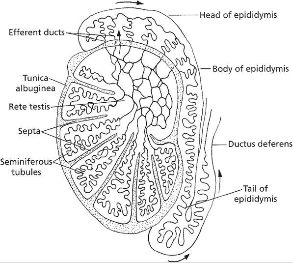
Figure 24-2. Internal anatomy of the testis. (Reprinted with permission of Wiley-Blackwell from Reece, W.O.
Functional Anatomy and Physiology of Domestic Animals, 3rd ed. Baltimore: Lippincott, Williams & Wilkins, 2005.)
serosa-lined cavity, carrying its serosal investments with it (discussed later).
Each testis consists of a mass of coiled seminiferous tubules (Fig. 24-2) surrounded by a heavy fibrous capsule called the tunica albuginea. A number of fibrous septa, also called trabeculae, pass inward from the tunica albuginea, dividing the testis into lobules and providing a framework for support of the seminiferous tubules and the interstitial tissue that produces testosterone. The seminiferous tubules are the site of spermatogenesis, the formation of spermatozoa.
The many seminiferous tubules deliver sperm into a network of tubules, the rete testis, which drains into the efferent ductules. The efferent ductules coalesce into a single epididymal duct.The connective tissue between the seminiferous tubules contains the interstitial cells (Leydig cells). The interstitial cells secrete the male hormone testosterone when stimulated by the pituitary gonadotropin luteinizing hormone (LH) (see Chapters 12 and 25). Sustentacular cells (Sertoli cells) within the seminiferous tubules envelop developing spermatozoa and their precursors. The sustentacular cells nourish the developing sperm and mediate the effects of follicle-stimulating hormone (FSH) and testosterone on the germ cells (Figs. 24-3 and 24-4).
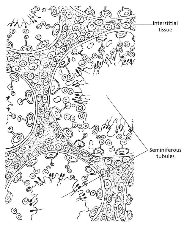
Figure 24-3. Illustration of seminiferous tubules surrounded by interstitial tissue. (Reprinted with permission of Wiley-Blackwell from Reece, W.O. Functional Anatomy and Physiology of Domestic Animals, 3rd ed. Baltimore: Lippincott, Williams & Wilkins, 2005.)
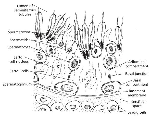
Figure 24-4. Cells of the seminiferous tubule. Sertoli cells surround and support developing spermatozoa. (Reprinted with permission of Wiley-Blackwell from Reece, W.O. Functional Anatomy and Physiology of Domestic Animals, 3rd ed. Baltimore: Lippincott, Williams & Wilkins, 2005.)