» The Abdominal Wall
The construction of the abdominal wall follows the common pattern in its essential features. The cutaneous muscle of the trunk is extensive as well as thick ventrally where it passes through the flank fold.
It leaves the abdominal floor uncovered, except for cranial (and inconstant caudal) preputial muscles. The deep fascia is without the elastic component that in the larger species imparts the characteristic yellow color. The three muscles of the flank show few distinctions of importance. Because the fleshy parts of the three flank muscles tend not to hold sutures well, the favorite site for laparotomy is an almost wholly tendinous aponeurotic strip, about 10 cm long and barely 5 cm wide, situated along the lateral edge of the rectus muscle and deep to the flank fold. The alternation of the abdominal muscles with layers of fat accounts for the characteristic appearance of the bacon rasher.Umbilical hernias used to be common in this species. If a satisfactory closure of these defects is to be obtained in the abdominal wall, it is first necessary to reflect the cranial part of the prepuce. This exposes the wide part of the linea alba that alone provides sufficient breadth of tissue to allow overlapping and suture of the margins of the hernia ring.
The other region of practical interest is provided by the inguinal canal. In principle, this conforms to the general arrangement of a potential space between the two oblique muscles (for details, see Fig. 34.2). The deep ring, the entry to the canal, is found between the caudal border of the internal oblique and the aponeurosis of the external oblique (see Fig. 2.27). The superficial opening is the split in the external aponeurosis that defines its division into pelvic and abdominal parts. The caudal part of the canal is very short, but it widens cranially because of the craniodorsal orientation of the deep ring compared to the slightly ventral and cranial angle of the superficial ring.
Anomalies of gubernacular development are common in pigs. If the canal is dilated (Fig. 34.3), pigs are predisposed to inguinal hernia involving a loop of small intestine that stretches the vaginal ring and forces a passage into the tunica vaginalis, leading to a subcutaneous swelling between the thighs. These hernias require attention in the castration of affected animals.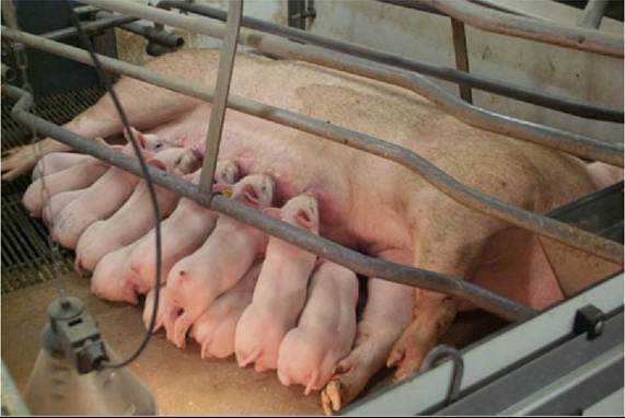
FIG. 34.1 The mammary glands of the sow extend from the pectoral to the inguinal region.
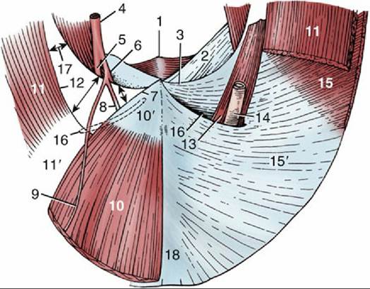
FIG. 34.2 Inguinal canal of the male made visible on the interior surface of the caudal abdominal wall; semi-schematic, cranial view. 1, Pelvic symphysis; 2, prepubic tendon; 3, caudal border of external oblique aponeurosis (“inguinal ligament”); 4, external iliac artery; 5, femoral artery; 6, deep femoral artery;
7, lateral border of rectus tendon; 8, external pudendal artery; 9, caudal epigastric artery; 10, rectus abdominis; 10', rectus tendon; 11, muscular part of internal abdominal oblique; 11', aponeurotic part of internal abdominal oblique; 12, caudal free border of internal abdominal oblique; 13, cremaster; 14, tunica vaginalis and spermatic cord; 15, muscular part of external ab-dominal oblique; 15', aponeurotic part of external abdominal oblique; 16, superficial inguinal ring; 17, deep inguinal ring (arrows); 18, linea alba.