THE ADRENAL GLANDS
The paired adrenal glands lie against the roof of the abdomen near the thoracolumbar junction. They are retroperitoneal and usually located craniomedial to the corresponding kidney (more directly medial in the horse).
Although they obtain their name from this relationship, they are in fact more closely connected with the major vessels in the abdomen—the aorta on the left, caudal vena cava on the right—and they adhere to these when the kidneys shift from the accustomed positions (e.g., the left kidney of the ruminant; see p. 693).Although generally elongated, the glands are often asymmetrical and quite irregular, being molded on neighboring vessels (Figure 6-6/1). It is difficult to specify their size because this appears to be influenced by several factors; they are relatively larger in wild than in related domestic forms, in juvenile than in adult individuals, and in pregnant and lactating females than in those reproductively inactive. Those of a medium-sized dog commonly measure about 2.5 ? 1 ? 0.5 cm.
Adrenal glands are firm, solid bodies that fracture readily when flexed. The fractured (or sectioned) surface exposes the division of the interior into an outer cortex and an inner medulla. The cortex, covered by a fibrous capsule, is yellowish and radially striated; the much
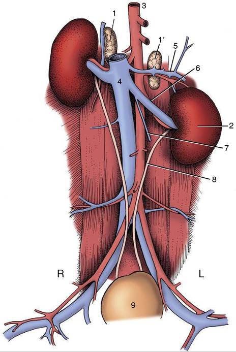
Figure 6-6 The topography of the canine adrenal glands. 1, 1', Right and left adrenal glands; 2, left kidney; 3, aorta; 4, caudal vena cava; 5, phrenicoabdominal vessels; 6, renal vessels; 7, ovarian vein; 8, ureter; 9, bladder.
darker medulla has a more uniform appearance. The two parts also contrast in origin, in microscopic structure, and in function.
The cortex is mesodermal and derived from a patch of celomic epithelium close to the gonadal fold.
On gross inspection, certain color changes vaguely suggest a subdivision into several concentric shells (zones), but these distinctions become clear only in microscopic preparations. The outer zone produces the mineralocorticoid hormone. The subjacent zones produce glucocorticoids and certain sex steroids.The medulla is of ectodermal origin, being contributed by a parcel of the cells that migrates from the neural crest to provide the neurons of the peripheral sympathetic ganglia. The medullary cells produce the transmitter substances norepinephrine and epinephrine and thus share with the sympathetic nervous system in the control of the body’s response (“flight or fight”) to acute stress situations. These cells obtain the additional designation chromaffin from their marked affinity for the salts of chromium and other heavy metals.
The adrenal glands are variously but always generously vascularized by small branches from several neighboring trunks: the aorta and the renal, lumbar, phrenicoabdominal, and cranial mesenteric arteries. After perfusing the gland, the blood pools within a central vein from which emissary vessels lead through a hilus to join the caudal vena cava or a tributary. Though not easily found, fine nerves within the cortex subject the tissue to hypothalamic control. Nerve bundles are more readily demonstrated within the medulla; appropriately, these are predominantly sympathetic preganglionic fibers passing to the medullary cells, which are equivalent to sympathetic postganglionic neurons elsewhere.
Accessory masses of cortical and medullary tissue both occur. Those of cortical tissue may be incorporated within any of several organs but are most commonly found attached to the capsule of the adrenal gland itself. Accessory chromaffin cells form the bodies known as paraganglia, which are endocrine cell clusters particularly associated with sympathetic nerves; a prominent example is found within the plexus on the aorta, close to the origin of the cranial mesenteric artery. Similar clumps of nonchromaffin cells, usually assigned to the parasympathetic system, are best known from the carotid and aortic bodies (described in Chapter 7, p. 241).