» The Anatomy of Rectal Exploration
Rectal palpation is possible in sows weighing 150 kg or more without great difficulty or ill effects on the animal. It is generally found that the small diameter and short suspension of the descending colon are greater impediments to these examinations than constriction of the pelvic canal.
With ample lubrication and sufficient cooperation, the arm can be introduced almost to the elbow; however, because the forearm is solidly wedged in the pelvic canal, the scope for exploration depends entirely on the length of and the mobility that may be exercised by the hand. The procedure allows examination of the pelvic inlet and bladder and, more important, the ovaries, cervix, and uterine artery for pregnancy diagnosis. The right kidney and the spiral colon- recognized through its coarse, granular content-may also be identified; the colon prevents access to the left kidney. Examination of the more confined pelvic cavity of boars is not feasible; the intrusion causes obvious pain.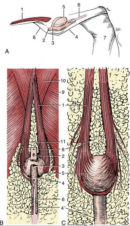
FIG. 35.10 Prepuce and preputial diverticulum. (A) In situ, Craniolateral view (schematic). (B) Ventral view. (C) Dorsal view. 1, Cranial preputial muscle, in (A) cut at both ends; 2, preputial orifice; 3, orifice
between prepuce and diverticulum; 4 and 4', wide cranial and narrow caudal parts of preputial cavity, respectively; 5, preputial diverticulum; 6, penis; 7, medial surface of right hock; 8, umbilicus; 9, cutaneous trunci; 10, pectoralis profundus; 11, preputial fat.
Comprehension Check
Compare the anatomic features of the male and female reproductive systems of the pig with those of the horse.
Review the blood and nerve supply to the pelvic viscera of the pig.