» The Anatomy of Rectal Palpation in Cattle*
As in the horse, rectal exploration in cattle is not free from risk of injury to the mucosa or even, in extreme cases, perforation of the intestinal wall—a mishap most likely to occur when invasion of the rectum induces straining.
Although rectal examinations of cows are most frequently performed to determine the health of the reproductive organs, it is also used to appreciate a much larger anatomic area once the hand is carried into the descending colon.The parts of the pelvic and abdominal walls that are accessible include the bones bounding the pelvic cavity and the regions of the deep inguinal rings. Dorsally, the caudal segment of the aorta and its bifurcation are within reach, and, scattered about the vessels, the larger lymph nodes of the medial iliac and deep inguinal groups (Fig. 29.4/13 and 14) can be palpated. The deep inguinal nodes are particularly important in connection with mastitis. The caudal part of the rumen is very obvious directly before the pelvic inlet, and it can be confirmed that ventrally the rumen extends into the right half of the abdomen. The caudodorsal blind sac may even intrude into the pelvic cavity when distended with gas. However, much of the rumen and the remaining compartments of the stomach are inaccessible, as are the liver and the spleen. The one necessary qualification of this statement refers to the abomasum, part of which is brought into reach in certain displacements. The right dorsal quadrant of the abdomen is occupied by small intestine, cecum, and colon, which together form a soft, fluctuating mass in which individual parts are mostly not identifiable when normal; the most common exception is the rounded tip of the gas-filled cecum.
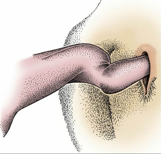
FIG. 29.36 Spiraling of the free part of the bovine penis in full erection.
Most of the left kidney, pushed to the right by the rumen and suspended from the abdominal roof, may be palpated; only the caudal pole of the right kidney is within reach and then only in smaller subjects. Healthy ureters are not detectable unless the initial portion of the left one can be appreciated where it passes over the surface of the kidney. The impression made by the bladder varies greatly because it forms a firm mass over the most cranial part of the pelvic floor when contracted but extends well forward into the abdomen as a fluctuating structure when distended. The intervention of the female reproductive tract makes this organ far less accessible in cows than in male animals.
A systematic examination of the reproductive tract is best begun by locating the cervix, easily recognized by its firmness and dimensions, although its location varies greatly according to the present status and past history of the animal. The short body of the uterus lies forward of the cervix, and the uterus may be fixed by the insertion of a finger between the intercornual ligaments to allow examination and comparison of the horns that diverge to each side. Frequently, these manipulations stimulate contraction of the uterine muscle, which can sometimes be quite powerful. The uterus may pass into the abdomen. If not too much enlarged, it may be retrieved by passing the hand forward and downward into the ventral part of the abdomen on the right side and then withdrawing the hand with the fingers flexed toward the palm to enclose the uterus. The broad ligaments proceeding to the horns of the uterus are distinct, but the uterine tubes, which run near the free cranial margins of the ligaments, are less certainly discoverable because, although fairly firm, they are only about 2 mm wide. The free margins of the broad ligaments also provide a guide to the location of the ovaries, which lie on the floor of the pelvic cavity in the young virgin animal but are displaced cranially and ventrally into the abdomen in older, more sexually experienced cows. An indication has already been given of the features of the follicles and corpora lutea that may be appreciated by examination of the ovarian surface. The reader is also reminded that the forward and downward movement of the reproductive organs in pregnancy may carry them out of reach for a time (Fig. 29.37).