The Anatomy of the Neuromuscular Junction Is Specialized for One-Way Communication
Motor neurons that synapse on skeletal muscles have their cell bodies located within the central nervous system (CNS), either within the spinal cord or the brainstem. The axons of these motor neurons travel within peripheral nerves, out to
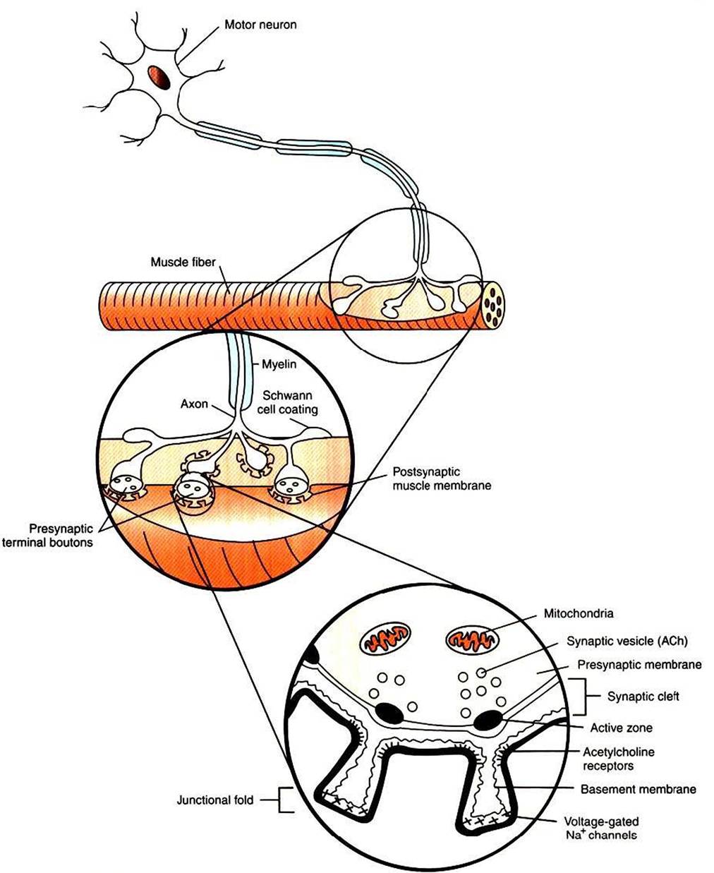
FIGURE 5-1 Synapse between a motor neuron and a skeletal muscle fiber.The neuromuscular junction has a presynaptic (neuronal) side, a narrow space between the neuron and muscle fiber called the synaptic cleft, and a postsynaptic (muscle) side.
ACh, Acetylcholine.the muscle» where each motor neuron synapses on several individual fibers (cells) of the muscle. However» an individual skeletal muscle fiber receives synaptic input from, and therefore its contraction is controlled by» only one motor neuron.
The neuromuscular junction, as with most chemical synapses, has a presynaptic side; a narrow space between the neuron and muscle fiber, called the synaptic cleft; and a postsynaptic side (see Figure 5-1). The presynaptic side of the synapse is made up of the terminal (transmitting) portion of the motor neuron. This presynaptic terminal has a swelled, buttonlike appearance and is also called a synaptic bouton. The terminal (or synaptic bouton) contains a large number of membranous storage vesicles, called synaptic vesicles, which contain the chemical neurotransmitter substance, in this case acetylcholine. These synaptic vesicles are lined up in rows along the inner surface of the terminal membrane (Figure 5-2). The presynaptic membrane region associated with each double row of vesicles is called an active zone and is the site where the synaptic vesicles will eventually release acetylcholine into the synaptic cleft. The presynaptic terminal also contains mitochondria, an indication of active metabolism in the cytoplasm.
Some mitochondrial products (e.g., acetyl-CoA, ATP) play a role in the local synthesis of acetylcholine and in its movement into the synaptic vesicles.The presynaptic (neural) and postsynaptic (muscle) cell membranes are separated by a narrow space, the synaptic cleft, that is about 50 nm wide (see Figures 5-1 and 5-2). The cleft contains extracellular fluid and a basal lamina, composed of a matrix of molecules, that is a specialized region of the muscle basement membrane. Some of these matrix molecules mediate synaptic adhesion between neuron and muscle.
The postsynaptic muscle cell membrane has several specialized features that facilitate synaptic transmission. Directly opposite the face of the presynaptic terminal, the postsynaptic muscle cell membrane contains receptors for the acetylcholine transmitter (see Figures 5-1 and 5-2). In this focal region the membrane has a series of invaginations, called junctional folds, that increase the surface area where acetylcholine receptors can reside. The acetylcholine receptors are most densely packed at the mouth of these junctional folds, and these mouths are closely aligned with the active zones of the presynaptic terminals from which the acetylcholine is released. Thus, there is a good match between the focal region of transmitter release from the neuron and the focal location of the receptors on the muscle fiber. Because the neurotransmitter is found only on the presynaptic neural side of the neuromuscular junction, transmission can go only from neuron to muscle, not in the reverse direction. Also, it should be noted that a motor neuron gives off several presynaptic terminals (synaptic boutons) to an individual muscle fiber. Together, this cluster of terminals is localized to a restricted region of the muscle fiber.
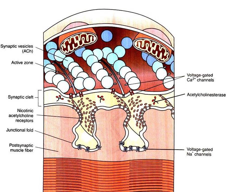
FIGURE 5-2 Presynaptic acetylcholine-filled synaptic vesicles line up at active zones, near voltage-gated Ca2* channels. Released acetylcholine binds with nicotinic acetylcholine receptors at junctional folds on the postsynaptic muscle fiber membrane (Redrawn and modified from Bear MF, Connors BW, Paradiso MA: Neuroscience: exploring the brain, ed 3, Philadelphia, 2007, LippincottWiIIiams & Wilkins.)