The Appendicular Skeleton
The appendicular skeleton is greatly modified by the conversion of the forelimbs to wings and by the hindlimbs assuming the sole responsibility for locomotion on the ground, perching, and withstanding the stresses of landing.
Avian long bones have thin, brittle cortices, unsuitable for the bone plating or pinning that might be contemplated for fracture repair in large cage birds. The bones of the forelimbs are braced against the axial skeleton, notably the sternum, by a well- developed shoulder girdle; the distal bones of the wing have undergone reduction. The skeleton of the hindlimb is strong and distally simplified by fusion and loss. In general, the pelvic limb is not involved in flight and is used more for swimming, catching prey, and wading.The Forelimb
The scapula (Fig. 37.9/23) is a flat rod lying lateral and parallel to the vertebral column and extending caudally to the pelvis. It is joined to the axial skeleton by muscles and ligaments. The cranial articulation with the clavicle receives the head of the humerus (shoulder joint). The strong coracoid bone (Fig. 37.9/24) extends from the shoulder joint to a firm articulation with the cranial end of the sternum and acts as a brace against the vigorous up-and-down strokes of the wing. The right and left clavicles unite to form the furcula (wishbone; Fig. 37.9/25), whose borders and median ventral expansion are tethered to the cranial end of the sternum and coracoids by tough membrane. The furcula connects the shoulder joints in springlike fashion and helps to brace the girdle against the axial skeleton. A foramen (canalis triosseus) at the junction of the scapula, coracoid, and clavicle transmits the tendon of one of the flight muscles.
The stout humerus (Fig. 37.9/9) is flat at both ends. The proximal extremity carries dorsal and ventral tubercles (Fig. 37.11). A pneumatic foramen (Fig.
37.11/4) is present close to the ventral tubercle. The ulna is thicker and longer than the radius (Fig. 37.11B and C). The proximal row of carpal bones is reduced by fusion to only two separate bones (radial and ulnar carpal bones; Fig. 37.11/6 and 7); the distal row has fused with the metacarpus. The number of metacarpal bones and corresponding digits is reduced to three.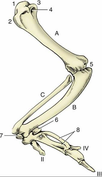
FIG. 37.11 Skeleton of the left wing, partially extended laterally; dorsal surface. 1, head; 2, dorsal tubercle; 3, ventral tubercle; 4, pneumatic foramen; 5, elbow joint; 6, ulnar carpal; 7, radial carpal; 8, Carpometacarpals; A, Humerus; B, ulna; C, radius; II to IV, digits.
The breast muscles that move the wing are well developed and in some species represent as much as 20% of the body weight. The pectoralis (Fig. 37.12/1), the superficial muscle, arises from the keel of the sternum and the clavicle and passes directly to the ventral surface of the dorsal tubercle of the humerus. Its contraction produces the powerful downbeat of the wing. The smaller Supracoracoideus (Fig. 37.12/2) also arises from sternum and clavicle. Its tendon is directed dorsally through the canalis triosseus and then across the head of the humerus to end close to its antagonist, the pectoralis. This muscle is used mainly for takeoff and is not employed in flight. The breast muscles are routinely palpated for an indication of the general health and condition of the bird. They are also used for intramuscular injection when care must be taken not to enter the body cavity (see Fig. 37.24/2 and 2'). However, pectoral injections should be avoided in birds relying on 100% flight efficiency: for example, birds of prey, homing pigeons, and wild birds due to be released. The cranial portion of the muscles should be avoided for this purpose because the larger vessels enter here and, if injured, may give rise to fatal hemorrhage. When intramuscular injections are given, the needle should be directed cranially, parallel to the sternum, to avoid puncture of the liver.
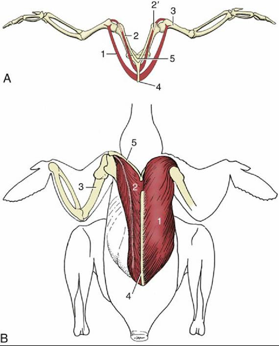
FIG.
37.12 Flight muscles. (A) Cranial view (schematic). (B) Dissected, ventral view (schematic). 1, Pectoralis; 2, supracoracoideus; 2', canalis triosseus for tendon of supracoracoideus; 3, humerus; 4, sternum; 5, clavicle.Pinioning is section of the tendon of the extensor carpi radialis at the carpal level that renders a bird unable to fly. This prominent muscle lies dorsal to the radius in the laterally extended wing. Its short tendon passes subcutaneously over the craniodorsal surface of the carpal joint and ends on the proximal end of the carpometacarpal bone (Fig. 37.13).
The propatagium, a triangular fold of skin, extends from the shoulder to the carpus and forms the leading edge of the wing. With the feathers, it is essential for producing aerodynamic lift, and tears or injuries to the propatagial ligament render the bird incapable of flight. These wounds are very difficult to repair.
The Hindlimb
The femur (Fig. 37.9B/21) resembles the mammalian bone in its general form. Its palpable proximal end may be used for sampling bone marrow. It slopes cranially (almost horizontally) to ensure that the large feet lie under the bird's center of gravity. A patella is present. The fusion of the tibia with tarsal elements forms a tibiotarsus (Fig. 37.9/31) that is much longer than the femur and carries the shaft of the feeble fibula on its lateral aspect. As in mammals, the knee joint has two menisci, cranial and caudal cruciate ligaments, and collateral ligaments. The fibula is robust proximally, where it articulates with the femur as well as the tibiotarsus, but is incomplete distally, tapering to a needlesharp point about three-quarters of the length down the tibiotarsus. This part of the limb is popularly known as the "drumstick." The distal tarsal elements merge with the metatarsal bone (itself a fusion of metatarsals II, III, and IV) to form the tarsometatarsus (Fig. 37.9/33). With no free tarsal bones present, the hock is an intertarsal joint with mainly flexion and extension movement.
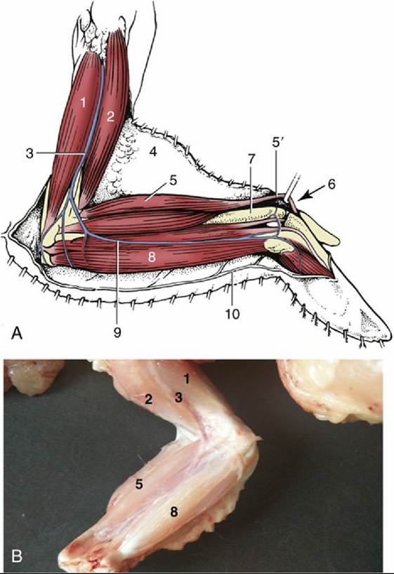
FIG. 37.13 (A) Laterally extended left wing, ventral surface (schematic). (B) Superficial dissection of laterally extended right wing, ventral surface. 1, Triceps; 2, biceps; 3, brachial vein; 4, skinfold (propatagium); 5, extensor carpi radialis; 5', tendon of extensor carpi radialis; 6, carpal joint; 7, subcutaneous part of radius; 8, flexor carpi ulnaris; 9, cutaneous ulnar (wing) vein; 10, reflected skin.
The Larsometatarsus extends to the ground, where it gives rise to four digits, although the phalangeal formula varies between species (Fig. 37.9A).
The caudal surface of the intertarsal joint bears a (tibial) cartilage through which the tendons of the digital flexors pass. The palpable gastrocnemius tendon passes through a sleeve connected to the caudal surface of the cartilage and ends on the plantar aspect of the tarsometatarsus. In the case of dietary insufficiency (perosis), which disfigures the cartilage, the tendons may slip off the hock and cause severe lameness and deformity. The digital flexors are arranged so that perching is possible with a minimum of muscular energy; lowering the body flexes knee and hock joints, which passively tenses the tendons that clamp the digits about the perch. The grip of a large bird can be undone by first extending the legs to relax the flexor tendons (Fig. 37.9A). Tendons of limb muscles generally ossify in large birds and become visible radiographically.
Red and white muscles (dark and white meat) are very clearly distinguished in birds. Red muscles contain larger amounts of myoglobin, are more heavily vascularized, and have more mitochondria and lipid globules within their fibers. They use fat rather than glycogen (carbohydrates) as a source of energy. Because fat supplies more energy than do carbohydrates per unit weight, muscles containing a predominance of red fibers are better suited to sustained effort. White muscles are more powerful but have less endurance. The breast muscles of birds with well-developed capacities for flight are red, those of the chicken and turkey are white, reflective of the galliform's preference for running. Selective breeding of farm-raised turkeys has greatly increased their weight and produced massive breast muscles.