THE ARTERIES
The arterial wall is composed of three concentric tunics (Figure 7-28). The endothelium of the inner one (tunica interna) is supported by a thin layer of specialized connective tissue that is bounded externally by a well- developed, fenestrated elastic sheet, the inner elastic membrane (Figure 7-28/2).
The subendothelial connective tissue is frequently affected by arteriosclerotic changes (hardening of the arteries), particularly, though not exclusively, in human subjects. The middle tunic (tunica media) is the thickest and most variable layer. It is composed of an elaborately organized admixture of elastic tissue and smooth muscle in varying proportions (Figure 7-28/3). The outer tunic (tunica adventitia) is predominantly fibrous and grades into the fibroareolar tissue within which many arteries are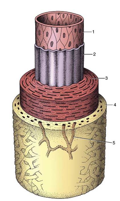
Figure 7-28 The components of the arterial wall. 1, 2, Tunica interna (1, endothelium; 2, inner elastic membrane); 3, tunica media; 4, tunica adventitia; 5, vasa vasorum.
embedded (Figure 7-28/4). Its importance in limiting expansion of the artery, which safeguards against spontaneous rupture, is not always sufficiently recognized.
Differences in the structure of the media allow the convenient recognition of three major classes of arteries, although it should not be assumed that these are sharply distinguished. A few very large arteries—those that are required to expand considerably when they receive the systolic output of the ventricles—have a media predominantly composed of concentric, fenestrated elastic membranes with relatively little muscle interspersed. The elastic tissue stretches to absorb and store the energy contained in the moving bloodstream; later, on recoil, it releases this energy to forward the flow of blood toward the periphery.
These elastic or conducting arteries comprise the first part of the aorta, certain of its major branches, and the pulmonary trunk.Most named arteries and others of smaller size have a media that consists largely of smooth muscle arranged in many closely spiraled layers. The caliber of these
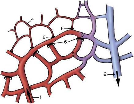
Figure 7-29 Schematic drawing of a capillary plexus. 1, Arteriole; 2, venule; 3, communicating (low-resistance) channel; 4, closed capillaries; 5, open capillaries; 6, precapillary sphincters.
muscular or distributing arteries is closely controlled by an autonomic innervation.
The smallest arteries, known as arterioles, principally regulate the resistance to the flow of blood and hence the peripheral blood pressure. The muscle is reduced to a few layers that are progressively shed. Although arterioles may be little wider than the capillaries into which they open, they are distinguished from these by the retention of some muscle in their walls. The sphincters about the openings to the capillaries are the means of determining the fraction of the capillary bed that is open to perfusion at any time (Figure 7-29).