THE ARTERIES
The first part of the aorta gives rise to right and left coronary arteries and a brachiocephalic trunk that immediately divides into right and left brachiocephalic arteries that send common carotid arteries forward into the neck and subclavian arteries toward the wings (Figure 37-16/'8",8"r').
In the thoracic inlet, the common carotids continue as internal carotids lying side by side on the ventral surface of the cervical vertebrae (Figure 37-15). The subclavian artery gives off a large pectoral trunk for the breast muscles and sternum before accompanying the humerus into the wing. In its descent along the vertebral column, the aorta gives rise to the following major branches: celiac (stomach, spleen, liver, intestine [Figure 37-29/2]), cranial mesenteric (intestines [Figure 37-29/5]), cranial renal (kidney, gonad [Figure 37-29/5]), external iliac (thigh [Figure 3729/12]), ischial (kidney, oviduct, hindlimb [Figure 3729/18]), and caudal mesenteric (intestine, cloaca). It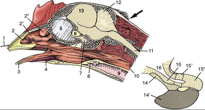
Figure 37-38 Median section of the head with an enlargement of the hypophysis (inset). The arrow indicates the approach to the foramen magnum through which euthanasia may be performed by injection into the brain. 1, Wire in nostril; 2,2', 2", rostral, middle, and caudal nasal conchae; 3, mandible; 4, tongue; 5, interorbital septum; 6, optic chiasm; 7, hypophysis (see also inset); 8, larynx; 9, trachea; 10, esophagus; 11, spinal cord; 12, cerebellum; 13, cerebrum; 14, 14', pars tuberalis and pars distalis of the adenohypophysis; 15, 15', 15", median eminence, infundibulum, and neural lobe of the neurohypophysis; 16, third ventricle.
ends by supplying the end of the oviduct, pelvic structures, and tail.