The Arteries
The arterial wall is composed of three concentric tunics (Fig. 7.29A and B). The endothelium of the inner one (tunica interna) is supported by a thin layer of specialized connective tissue that is bounded externally by a well-developed, fenestrated elastic sheet, the inner elastic membrane (Fig.
7.29/2). The subendothelial connective tissue is frequently affected by arteriosclerotic changes (hardening of the arteries), particularly though not exclusively in humans. The middle tunic (tunica media) is the thickest and most variable layer. It is composed of an elaborately organized admixture of elastic tissue and smooth muscle in varying proportions (Fig. 7.29/3). The outer tunic (tunica adventitia) is predominantly fibrous and grades into the fibroareolar tissue that limits expansion of the artery (Fig. 7.29/4).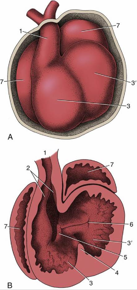
FIG. 7.28 The partitioning of the truncus arteriosus. (A) Ventral view of the developing heart. (B) The ventral part of the heart has been removed to expose the developing ridges (2) in the truncus arteriosus. 1, Truncus arteriosus; 2, ridges in truncus; 3, right ventricle; 3', left ventricle; 4, interventricular septum; 5, right atrioventricular canal; 6, left atrioventricular canal; 7, atrium.
The differences in the structure of the media results in three major classes of arteries. A few very large arteries that stretch upon receiving the systolic output of the ventricles have a media composed predominantly of concentric, fenestrated elastic membranes with relatively little muscle interspersed. The elastic tissue stretches to absorb and store the energy followed by its release upon recoil to aid the flow of blood toward the periphery. These elastic or conducting arteries are the first part of the aorta, certain of its major branches, and the pulmonary trunk.
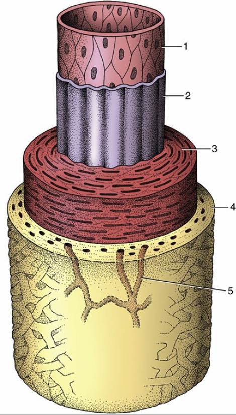
FIG. 7.29 The components of the arterial wall. 1 and 2, Tunica interna (1, endothelium; 2, inner elastic membrane); 3, tunica media; 4, tunica adventitia; 5, vasa vasorum.
The media of most named arteries and others of smaller size is composed largely of many closely spiraled layers of smooth muscles. The caliber of these muscular or distributing arteries is closely controlled by an autonomic innervation.
The smallest arteries, known as arterioles, principally regulate the resistance to the flow of blood and hence the peripheral blood pressure. The muscle, reduced to a few layers, is progressively shed. The openings of arterioles into capillaries are guarded by sphincters that regulate the vascular perfusion of capillary bed (Fig. 7.30).