» The Blood Vessels and Lymphatic Structures of the Hindlimb
The femoral artery continues the external iliac artery beyond the vascular lacuna. It passes between the medial muscles of the thigh to reach the flexor surface of the stifle, where it is renamed the popliteal artery.
This soon divides into cranial and caudal tibial arteries (Fig. 31.8/10 and 11). One branch of the femoral, the saphenous artery (Fig. 31.8/7), runs on the surface of the gracilis and is often used for taking the pulse of cows by sliding the hand from behind, between the udder and thigh. This vessel is responsible for the vascularization of the caudal part of the leg and follows the common calcanean tendon to the hock, where it gives rise to medial and lateral plantar arteries.The cranial tibial artery (Fig. 31.6/8), which may be regarded as the continuation of the femoral trunk, runs embedded between the crural muscles to reach the flexor (dorsal) surface of the hock joint under cover of the long digital extensor tendon. The caudal tibial artery is of minor local significance.
Renamed the dorsal metatarsal artery (Fig. 31.8/12), the main trunk now sends a perforating artery through the upper part of the metatarsal bone before continuing in the dorsal groove of this bone. A second perforating artery is released toward the fetlock. The perforating branches join the plantar arteries and are also connected by small deeper vessels. The plantar arteries resemble the corresponding forelimb vessels. One branch of the medial plantar artery crosses the plantar surface of the medial tendon of the superficial flexor proximal to the fetlock and is here liable to injury.
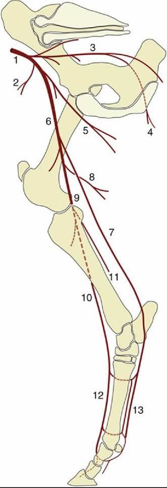
FIG. 31.8 The principal arteries of the bovine right hindlimb, medial view. 1, External iliac artery (a.); 2, deep circumflex iliac a.; 3, internal iliac a.; 4, caudal gluteal a.; 5, deep femoral a.; 6, femoral a.; 7, saphenous a.; 8, caudal femoral a.; 9, popliteal a.; 10, cranial tibial a.; 11, caudal tibial a.; 12, dorsal metatarsal arteries; 13, medial and lateral plantar and metatarsal (closer to the bone) arteries.
This branch continues into the interdigital space, where it anastomoses with the main trunk. The anastomosis is substantial and winds around below the proximal interdigital ligament, where it is encountered in amputation of a digit. The axial surfaces of the digits are supplied by branches arising from the anastomosis. The abaxial surfaces are supplied by direct continuations of the plantar arteries. There are a large number of other anastomoses that do not merit description here.
The veins are divided between a deep system satellite to the arteries and a few superficial vessels that follow independent courses (Fig. 31.9). The superficial vessels comprise the medial and lateral saphenous veins and their tributaries. The larger lateral saphenous vein (Fig. 31.9A/9) arises from two tributaries: one ascends with the extensor tendons and superficial peroneal nerve and crosses on the dorsolateral aspect of the hock, and the other ascends with the lateral plantar artery from a subcutaneous origin on the lateral digit and follows the flexor tendons under cover of the deep fascia to cross the joint plantarolaterally. The lateral saphenous vein raises a ridge below the skin as it crosses to the caudal border of the leg and then follows the curvature of the gastrocnemius, eventually to open into the femoral vein. The medial saphenous vein (Fig. 31.9A/5) is also formed by two tributaries. The more important caudal one takes its origin from the abaxial aspect of the medial digit, ascends with the medial plantar artery, and passes the hock plantaromedially. The medial saphenous vein ascends together with the palpable saphenous artery on the medial aspect of the leg to dip between the gracilis and sartorius muscles to join the femoral vein.
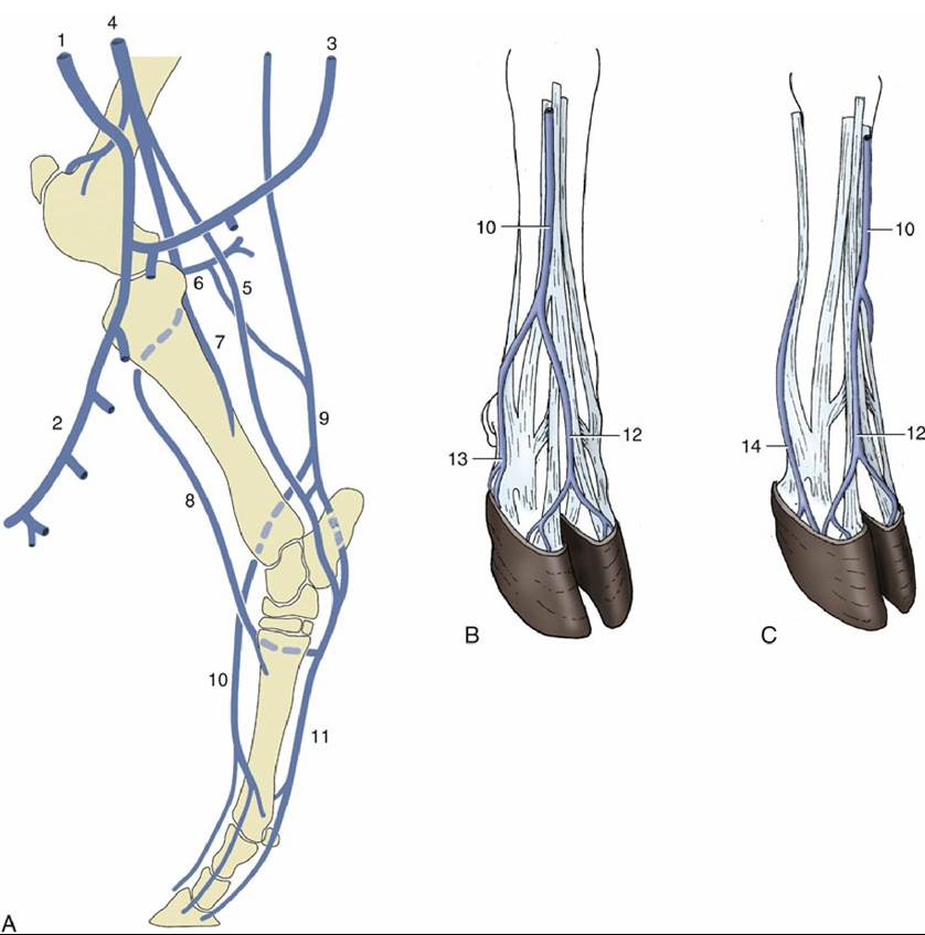
FIG. 31.9 The major veins of the bovine hindlimb. (A) Right limb, medial view. (B) Right hindfoot, dorsolateral view. (C) Left hindfoot, dorsomedial view.
1, External pudendal vein (v.); 2, mammary v.; 3, ventral labial v.; 4, femoral v.; 5, medial saphenous v.; 6, caudal femoral v.; 7, caudal tibial v.; 8, cranial tibial v.; 9, lateral saphenous v.; 10, cranial tributary of lateral saphenous v.; 11, medial and lateral plantar veins; 12, dorsal common digital v. III; 13, plantar v. of lateral digit; 14, plantar v. of medial digit.The superficial veins (Fig. 31.9B and C) may be raised by application of a tourniquet below the hock for injection of local anesthetic so that the digits may be desensitized.
The lymph nodes include the popliteal node within the popliteal fossa and the very large subiliac node described with the abdominal wall (Fig. 31.10/9 and 10). A small coxal node ventral to the coxal tuber and a group of gluteal nodes on the lateral surface of the sacrosciatic ligament are also commonly present (Fig. 31.10/2 and 5). An ischial node (Fig. 31.10/6) that lies on the ligament just dorsal to the lesser sciatic foramen can be inspected in the split carcass by incision of the ligament from within the pelvis. A tuberal node (Fig. 31.10/7) lies medial to the ischial tuber within the ischiorectal fossa.
The popliteal node collects from the distal part of the limb, including most of the leg, and sends its efferent vessels along two routes: one follows the sciatic nerve to the ischial node, and the second accompanies the femoral vessels to the large, deep inguinal node (Fig. 31.10/4) at the side of the pelvic inlet. The subiliac node drains the skin over the thigh and stifle in addition to the flank; its efferents also go chiefly to the deep inguinal node. The smaller nodes are only of local significance.