The brachial plexus is formed by the last three cervical and first two thoracic nerves.
Its branches generally conform to the common pattern, but some points merit repetition or amplification because of their clinical relevance.
The suprascapular (C6-C7) nerve winds around the cranial border of the scapula to reach the supraspinatus and infraspinatus muscles (Fig.
30.15). Destruction has little effect on the standing posture beyond producing occasional slight abduction of the arm. Walking is more severely affected, and the limb is advanced with a stiff, circumducted stride while the shoulder is abducted most obviously in the support phase. In chronic paralysis the muscles atrophy and the scapular spine becomes sharply defined.The large median nerve (C8-T2) runs down the medial aspect of the arm, crosses the elbow joint (where it is palpable in front of the brachial artery), and dips under the flexor muscles to which it sends branches. The much-reduced trunk then follows the median artery under cover of the flexor carpi radialis (Fig. 30.3/2) into the carpal canal before dividing in midmetacarpus into several branches that supply most of the palmar aspect of the foot.
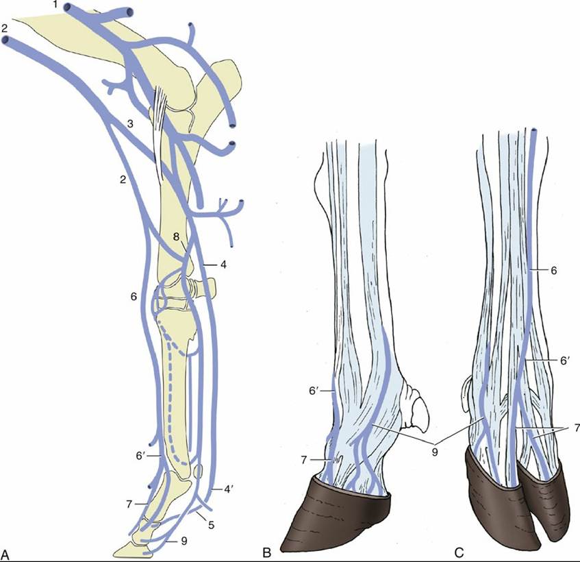
FIG. 30.14 The principal veins (vv.) of the bovine forelimb. (A) Right limb, medial view. (B) Left foot, lateral view. (C) Right foot, dorsal view. 1, Brachial vein (v.); 2, cephalic v.; 3, median cubital v.; 4, median v.; 4', palmar common digital v. III; 5, axial palmar digital vv.; 6, accessory cephalic v.; 6', dorsal common digital v. III; 7, dorsal digital vv.; 8, radial v.; 9, abaxial palmar digital vv.
The ulnar nerve (C8-T2) arises with the median nerve but diverges from this in midarm (Fig. 30.15/15). After releasing a branch to the skin, it passes toward the olecranon, where it dips between the origins of the flexor muscles. It detaches branches to these before continuing as a mainly sensory nerve (Fig.
30.3/4') that divides a short distance above the accessory carpal bone. The palmar branch runs through the carpal canal lateral to the flexor tendons. The dorsal branch becomes superficial and may be palpated where it descends over the lateral aspect of the accessory carpal bone.Because the median and ulnar nerves share in the supply of the carpal and digital flexors, destruction of either one has little effect on posture or gait. Even when both are sectioned, no immediate change in the appearance of the standing animal occurs, although overextension of the carpus develops later. Walking is affected by the double neurectomy and is performed with an exaggerated "goose-stepping" action in which the carpal and lower joints are overextended. However, the stride is not shortened, and the foot remains able to support weight.
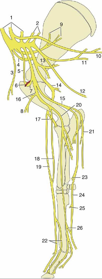
FIG. 30.15 Nerves of the bovine forelimb, medial view. 1 and 2, Roots of brachial plexus; 3, cranial pectoral nerve (n.); 4, suprascapular n.; 5, musculocutaneous n.; 6, axillary artery; 7, loop of musculocutaneous n. before joining median n.; 8, proximal branch of musculocutaneous n.; 9, subscapular n.; 10, long thoracic n.; 11, thoracodorsal n.; 12, lateral thoracic n.; 13, axillary n.; 14, radial n.; 15, ulnar n.; 16, combined musculocutaneous and median nerves; 17, distal branch of musculocutaneous n.; 18, medial cutaneous antebrachial n.; 19, superficial branch of radial n.; 20, median n.; 21, caudal cutaneous antebrachial n.; 22, dorsal common digital nerves III and II; 23, dorsal branch of ulnar n.; 24, palmar branch of ulnar n.; 25, deep branch of ulnar n. (to interosseous muscles); 26, communicating branch.
The radial nerve (C7-T1) lies more caudally in the arm. It dives between the heads of the triceps before following the brachialis to reach the cranial surface of the elbow while furnishing muscular branches en route.
The trunk is vulnerable as it passes over the sharp epicondyloid crest of the humerus deep to the lateral head of the triceps. In this position it divides into several branches that innervate the extensor muscles of the carpus and digits and a cutaneous branch that accompanies the cephalic and, more distally, the accessory cephalic vein. It is joined by a branch of the musculocutaneous nerve before crossing the carpus (Fig. 30.15/18 and 19). The radial nerve is the exclusive supply to the extensors of all joints distal to the shoulder, and the effects of injury in the proximal part of its course are correspondingly severe. The elbow is "dropped," and the limb appears to be abnormally long. The animal moves with difficulty, dragging the toes and taking no weight on the affected limb. It is unable to place the sole of the hoof on the ground and rests on the dorsal surfaces of the digits. If the damage is more distal, the animal can usually learn to compensate for the loss of carpal and digital extensor muscle function.Nerve-blocking procedures, such as are widely used in equine practice, are not used for the differential diagnosis of lameness in cattle. Because the retrograde intravenous techniques used to secure anesthesia for the performance of digital surgery are now so popular, it seems unnecessary to supply detailed accounts of the digital nerves, which are available in reference texts.
In very brief summary, it may be stated that the dorsal aspect of the foot is the province of the radial nerve, the palmar aspect is the province of the median nerve, and the lateral aspect the province of the ulnar nerve (Fig. 30.16A-C).