The Cellular Component of Blood Includes Red Blood CeIIsrWhite Blood Cells, and Platelets
Cells normally constitute 30% to 60% of the blood volume (depending on the species). The fraction of cells in blood is called the hematocrit (see Figure 18-5). The hematocrit is determined by adding an anticoagulant to some blood and then centrifuging it in a tube.
The cells are somewhat heavier than plasma and settle to the bottom of the tube during centrifugation. Because centrifugation results in a packing of the blood cells in the bottom of the tube, the hematocrit is sometimes called the packed cell volume. Most of the cell component looks red because most of the blood cells are erythrocytes (red blood cells, RBCs). Erylhrocytes acquire their red color from hemoglobin.The leukocytes (white blood cells, WBCs) are slightly lighter in weight than the RBCs; in a centrifuge tube the WBCs gather in a white “buffy coat” on top of the RBCs. The huffy coat is normally very thin because there are about IOOO times more RBCs than WBCs. Leukocytes are critical in immune and allergic responses of the body. The subtypes of leukocytes include neutrophils, lymphocytes, monocytes, eosinophils, and basophils. A laboratory analysis of the total number and relative distribution of the various WBC subtypes (differential white blood cell count) provides important clues in the diagnosis of disease. Both erythrocytes and leukocytes are made in the bone marrow. They develop, by mitosis and differentiation, from a common line of progenitor cells, the pluripotent (uncommitted) stem cells.
The cellular component in a centrifuge tube also contains platelets, or thrombocytes, which are fragments of the membranes of their precursor cells, the megakaryocytes. The megakaryocytes reside in the bone marrow and shed platelets into the bloodstream. Platelets participate in hemostasis (the control ofbleeding through coagulation and clotting). In this process a clumping together of platelets (platelet aggregation) begins to create a physical barrier across openings in blood vessels.
Substances released from the platelets, along with fibrinogen and several clotting factors in the plasma, lead to the coagulation of blood and the formation of a stable, fibrin-based blood clot.
Coagulation and clotting involve complex, interconnected sequences of chemical reactions (the coagulation cascade). A key step in the coagulation cascade is the formation in the plasma of thrombin, an enzyme that catalyzes the transformation of fibrinogen to fibrin. Several laboratory tests are used to assess the status of an animal’s coagulation system. The two most common tests involve determination of the prothrombin time (PT) and the partial thromboplastin time (PTT).
Ifblood is allowed to coagulate and then is centrifuged, the fibrin and other plasma clotting factors settle to the bottom along with the RBCs, WBCs, and platelets. The liquid portion remaining above (essentially plasma without fibrinogen and other clotting factors) is called serum. Most of the common clinical blood chemistry analyses are performed on serum. Examples include the determination of concentrations of electrolytes and cholesterol.
Ifblood is treated with an anticoagulant and then allowed simply to sit in a tube (without centrifugation), the erythrocytes slowly begin to settle. For reasons that are not completely understood, the rate of their settling tends to be increased to above normal in certain disease states and decreased to below normal in others. Therefore the erythrocyte sedimentation rate (ESR) is a clinically useful diagnostic measurement. The normal ESR varies substantially between species; for example, it is much more rapid in equine blood than in canine blood.
Blood cell counts are performed by manual or automated scanning of a very small volume (e.g., 1 μL) of anticoagulated whole blood. Table 18-4 presents a summary of normal hematologic values for the dog.
Table 18-4
Canine Hematology
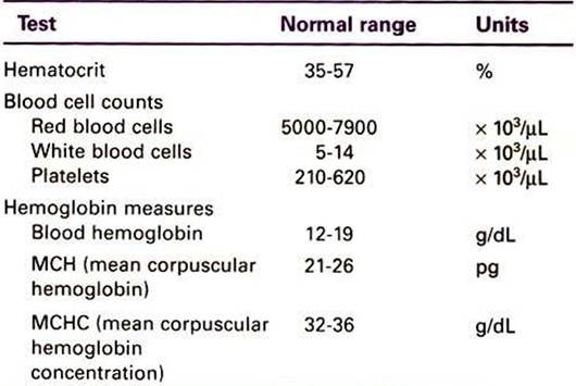
Modified from Latimer KSr Mahaffey EAr Prasse KW: Duncan & Prasse's veterinary laboratory medicine: clinical pathology, ed 4r Amesr 2003r Iowa State University Press.