The Central Nervous System Can Be Divided into SixAnatomicaI Regions
The CNS has a longitudinal organization, with the Phylogenetically oldest parts lying more caudal and the newest portions lying rostral. The CNS can be divided into six major regions (Figure 3-2): the spinal cord and five major brain regions.
From caudal to rostral, these brain regions are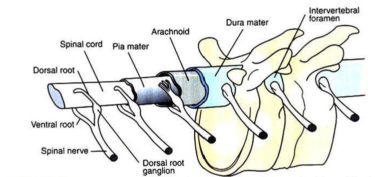
FIGURE 3-1 Spinal cord and the three layers of the meninges within the vertebral canal. Action potentials generated on sensory afferents enter the spinal cord along axons in the dorsal roots.Those generated on motor efferents exit the spinal cord along axons in the ventral roots. (Redrawn from Gardner E: Fundamentals of neurology, ed 3, Philadelphia, 1959, Saunders.)
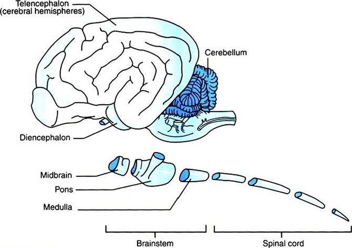
FIGURE 3-2 Central nervous system (CNS) has longitudinal organization in which the Phylogenetically oldest parts are caudal and the newest parts are rostral. The CNS can be divided into six major regions: the spinal cord, medulla, pons, midbrain, diencephalon, and telencephalon (cerebral hemispheres).
the medulla, pons, midbrain, diencephalon, and telencephalon. (The cerebellum, a brain structure that lies dorsal to portions of the pons and medulla, is sometimes named as a seventh major region of the CNS.) The medulla, pons, and midbrain form the brainstem; the diencephalon and telencephalon form the forebrain.
In general, the spinal cord, brainstem, and forebrain represent a hierarchy of functional organization. The spinal cord receives sensory input from and supplies motor output to the trunk and limbs; the brainstem performs these functions for the face and head. Sensory information entering the brainstem is passed to the forebrain, where the most sophisticated forms of information processing take place.
Sensory information entering the spinal cord is relayed to the forebrain by way of the brainstem. The forebrain also formulates the most sophisticated forms of motor output. This output is sent to the brainstem for executing movement of the face and head or for relay to the spinal cord to execute trunk and limb movement. The forebrain is also capable of sending motor commands directly to the spinal cord.Each of the six CNS regions has distinctive anatomical and functional characteristics. Some of these include the following:
1. The spinal cord is the most caudal region in the CNS. As previously noted, it receives action potentials along sensory dorsal root axons from receptors in the skin, muscles, tendons, joints, and visceral organs. It contains the cell bodies and dendrites of motor neurons whose axons exit through the ventral roots either to reach skeletal muscles or to reach out toward smooth muscle. The spinal cord also contains tracts of axons carrying sensory information to the brain and motor commands from the brain to the motor neurons. The isolated spinal cord can control simple reflexes, such as muscle stretch reflexes and limb withdrawal to painful stimuli.
2. The medulla lies rostral to the spinal cord and resembles it in many ways. By way of cranial nerves, the medulla too receives information from the body’s external and internal sensory receptors and sends motor commands out to skeletal and smooth muscle. Large populations of these receptors and muscles lie in the head and neck region. The cell bodies of medullary neurons that receive the sensory input from cranial nerves or that send the motor output are respectively collected in aggregates called sensory or motor cranial nerve nuclei. The cranial nerve nuclei of the medulla play a critical role in life support functions of the respiratory and cardiovascular systems and in aspects of feeding (e.g., taste, tongue movement, swallowing, digestion) and vocalization.
3. The pons lies rostral to the medulla and contains the cell bodies of large numbers of neurons in a two-neuron chain that relays information from the cerebral cortex to the cerebellum.
The cerebellum is not a part of the brainstem but is often described along with the pons because of a similar embryological origin. The cerebellum is important for smooth, accurate, coordinated movement and for motor learning. Cranial nerve nuclei of the pons play important roles in receiving sensory information about facial touch and in the motor control of chewing.4. The midbrain, or mesencephalon, lies rostral to the pons and contains the superior and inferior colliculi, which are important in processing and relaying visual and auditory information that has entered the brain at other levels. The midbrain also contains cranial nerve nuclei that directly control eye movement and regions that coordinate some eye movement reflexes.
Each region of the brainstem contains axon tracts carrying action potentials to or from the forebrain, as well as tracts that carry action potentials to or from the spinal cord. Each brainstem region also contains a portion of the reticular formation, a netlike complex of many small clusters of cell bodies (nuclei) and loosely organized axonal projections, located near the midline. The reticular formation plays important roles in modulating consciousness and arousal, pain perception, and spinal reflexes, as well as in movement.
5. The diencephalon contains the thalamus and the hypothalamus, both of which are large structures consisting of several subnuclei. The thalamus is a relay station for and a modulator of information being passed to the cerebral cortex from sensory systems and other brain regions. The hypothalamus regulates the autonomic nervous system, controls hormone secretion of the pituitary gland, and plays a major role in physiological and behavioral aspects of homeostasis (e.g., maintenance of temperature and blood pressure; feeding).
6. The telencephalon, also commonly referred to as the cerebral hemispheres, is made up of the cerebral cortex and a small number of prominent subcortical structures, such as the basal ganglia and hippocampus. The cerebral cortex mediates the most complex forms of sensory integration and conscious sensory perception. It also formulates and executes sequences of voluntary movement. The basal ganglia are a collection of nuclei that modulate the motor functions of cerebral cortex, and the hippocampus plays an important role in memory and spatial learning.