The Cochlea Transduces Sound Waves to Action Potentials of the Eighth Cranial Nerve
The organ of Corti mediates the transduction of sound waves into action potentials. The hair cell receptors of the organ of Corti are similar in structure and function to the hair cells that form the vestibular sensory organs.
The hair cells synapse on sensory neurons that form the cochlear portion of the eighth cranial (vestibulocochlear) nerve, which projects to the brainstem’s cochlear nuclei. The cell bodies of these sensory neurons reside in the spiral ganglion. Sound-induced bending of the hair cell cilia changes the frequency of action potentials on the eighth nerve fibers.Sound waves in the external environment are collected by the outer ear and cause vibrations of the tympanic membrane. These vibrations are transmitted through the middle ear by movement of the ossicles and result in similar vibrations of the oval window of the cochlea. As the oval window vibrates, sound energy is transferred down through the perilymph of the scala vestibuli and down through the endolymph of the scala media to the basilar membrane. This energy produces a series of traveling waves that begin near the base of the basilar membrane (closest to the oval window) and move along its length. The situation is analogous to whipping the
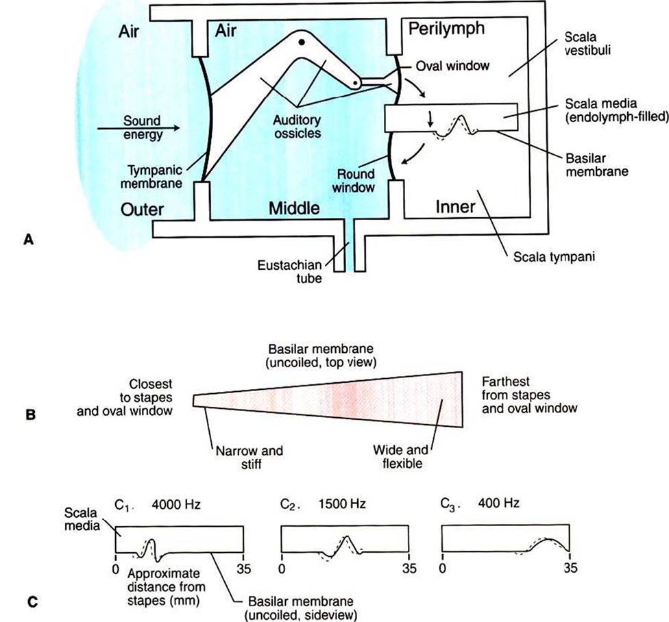
FIGURE 17-4 Schematic representation of the transmission of sound energy from the outer ear to the inner ear. A, Sound energy enters the air-filled outer ear and vibrates the tympanic membrane, which produces movement of the ossicles in the air-filled middle ear. Ossicular movement displaces the oval window of the fluid-filled inner ear, resulting in a traveling wave on the basilar membrane. B, Basilar membrane is narrow and stiff at the base and wider and more flexible farther from the base. C, As the frequency of a sound decreases, the region of maximum displacement of the basilar membrane, produced by the traveling wave, is located progressively farther from the base. (Redrawn from Lippold OCJ, Winton FR: Human physiology, ed 6, NewYork, 1972, Churchill Livingstone.)
free end of a rope that is stationary at the opposite end.
A diagram of this transmission is shown in Figure 17-4. The movement of the traveling wave causes portions of the flexible basilar membrane to move up and down. Because the organ of Corti sits atop the basilar membrane, this up-and-down motion causes the hair cell cilia to be sheared back and forth against the anchored, overlying tectorial membrane (Figure 17-5). This, in turn, changes the release of transmitter from the hair cells onto the eighth nerve neurons, which in turn alters the action potential firing rate of these neurons. It is at this point that the organ of Corti has transduced the sound wave energy into neural activity. As the amplitude of an environmental sound wave increases (normally perceived as a louder sound), a longer area of the basilar membrane is displaced. This results in a larger number of hair cells shearing against the tectorial membrane, which in turn affects the activity of a larger number of eighth nerve neurons. This is one way that sound intensity is coded by the nervous system.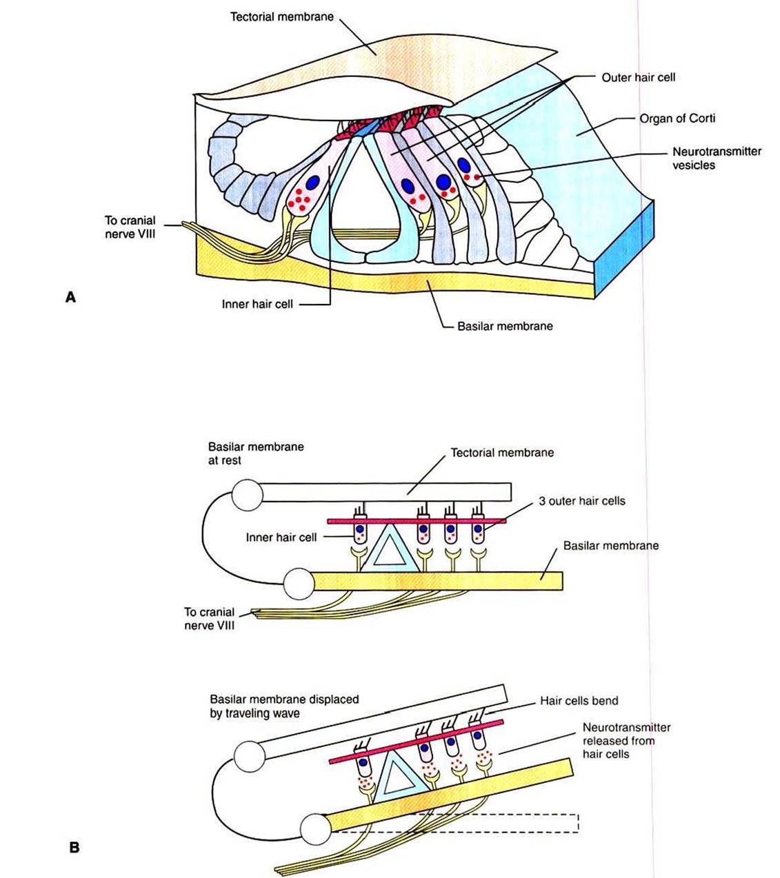
FIGURE 17-5 Transduction of sound into neural activity occurs at the organ of Corti. A, Structural organization of the organ of Corti and its hair cell receptors, sitting atop the basilar membrane. BzTraveIing waves along the basilar membrane displace the membrane and organ of Corti upward, shearing hair cell cilia against the overlying tectorial membrane. This bending of the cilia induces the release of neurotransmitter from the hair cells onto sensory neurons of the eighth cranial nerve. (Modified from Goldstein BE: Sensation and perception, ed 6, Pacific Grove, Calif, 2002, Wadsworth.)